9FCF

Medicago truncatula 5'-ProFAR isomerase (HISN3) D57N mutant in complex with ProFAR


Experimental Data Snapshot

  • Method: X-RAY DIFFRACTION
  • Resolution: 2.36 Å
  • R-Value Free: 0.257 
  • R-Value Work: 0.224 
  • R-Value Observed: 0.227 

Starting Model: in silico
View more details

wwPDB Validation   3D Report Full Report


Ligand Structure Quality Assessment 


This is version 1.0 of the entry. See complete history


Literature

Structural, kinetic, and evolutionary peculiarities of HISN3, a plant 5'-ProFAR isomerase.

Witek, W.Imiolczyk, B.Ruszkowski, M.

(2024) Plant Physiol Biochem 215: 109065-109065

  • DOI: https://doi.org/10.1016/j.plaphy.2024.109065
  • Primary Citation of Related Structures:  
    9FCF, 9FCG

  • PubMed Abstract: 

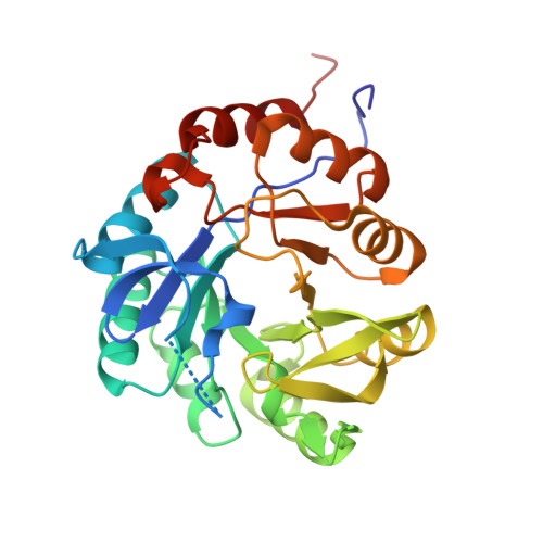
    Histidine biosynthesis is essential for the growth and development of plants, where it occurs within chloroplasts. The eleven reactions are catalyzed by eight enzymes, known as HISN1-8, each acting sequentially. Here, we present the crystal structures of a 5'-ProFAR isomerase (HISN3) from the model legume Medicago truncatula bound to its enzymatically synthesized substrate (ProFAR) and product (PrFAR). The active site of MtHISN3 contains a sodium cation that participates in ligand recognition, a feature not observed in bacterial and fungal structures of homologous enzymes. The steady-state kinetics of wild-type MtHISN3 revealed a slightly higher turnover rate compared to its bacterial homologs. Plant HISN3 sequences contain an unusually elongated Lys60-Ser91 fragment, while deletion of the 74-80 region resulted in a 30-fold loss in catalytic efficiency compared to the wild-type. Molecular dynamics simulations suggested that the fragment facilitates product release, thereby contributing to a higher k cat . Moreover, conservation analyses suggested a non-cyanobacterial origin for plant HISN3 enzymes, which is another instance of a non-cyanobacterial enzyme in the plant histidine biosynthetic pathway. Finally, a virtual screening campaign yielded five molecules, with the energy gains ranging between -13.6 and -13.1 kcal/mol, which provide new scaffolds for the future development of herbicides.


  • Organizational Affiliation

    Department of Structural Biology of Eukaryotes, Institute of Bioorganic Chemistry, Polish Academy of Sciences, Noskowskiego 12/14, 61-704, Poznan, Poland.


Macromolecules
Find similar proteins by:  (by identity cutoff)  |  3D Structure
Entity ID: 1
MoleculeChains Sequence LengthOrganismDetailsImage
1-(5-phosphoribosyl)-5-[(5-phosphoribosylamino)methylideneamino] imidazole-4-carboxamide isomerase, chloroplastic274Medicago truncatulaMutation(s): 1 
Gene Names: 11441829MTR_2g015010MtrunA17_Chr2g0283151
EC: 5.3.1.16
UniProt
Find proteins for G7IFI7 (Medicago truncatula)
Explore G7IFI7 
Go to UniProtKB:  G7IFI7
Entity Groups  
Sequence Clusters30% Identity50% Identity70% Identity90% Identity95% Identity100% Identity
UniProt GroupG7IFI7
Sequence Annotations
Expand
  • Reference Sequence
Small Molecules
Ligands 3 Unique
IDChains Name / Formula / InChI Key2D Diagram3D Interactions
GUO (Subject of Investigation/LOI)
Query on GUO

Download Ideal Coordinates CCD File 
B [auth A][(2R,3S,4R,5R)-5-[4-aminocarbonyl-5-[(E)-[[(2R,3R,4S,5R)-3,4-bis(oxidanyl)-5-(phosphonooxymethyl)oxolan-2-yl]amino]methylideneamino]imidazol-1-yl]-3,4-bis(oxidanyl)oxolan-2-yl]methyl dihydrogen phosphate
C15 H25 N5 O15 P2
QOUSHGMTBIIAHR-KEOHHSTQSA-N
CL
Query on CL

Download Ideal Coordinates CCD File 
D [auth A]CHLORIDE ION
Cl
VEXZGXHMUGYJMC-UHFFFAOYSA-M
NA
Query on NA

Download Ideal Coordinates CCD File 
C [auth A]SODIUM ION
Na
FKNQFGJONOIPTF-UHFFFAOYSA-N
Experimental Data & Validation

Experimental Data

  • Method: X-RAY DIFFRACTION
  • Resolution: 2.36 Å
  • R-Value Free: 0.257 
  • R-Value Work: 0.224 
  • R-Value Observed: 0.227 
  • Space Group: P 4
  • Diffraction Data: https://doi.org/10.18150/DGZKW3
Unit Cell:
Length ( Å )Angle ( ˚ )
a = 91.301α = 90
b = 91.301β = 90
c = 35.836γ = 90
Software Package:
Software NamePurpose
PHENIXrefinement
XDSdata scaling
PHASERphasing
Cootmodel building
XDSdata reduction

Structure Validation

View Full Validation Report


Ligand Structure Quality Assessment 


Entry History & Funding Information

Deposition Data


Funding OrganizationLocationGrant Number
Polish National Science CentrePolandSonata 2018/31/D/NZ1/03630

Revision History  (Full details and data files)

  • Version 1.0: 2024-09-04
    Type: Initial release