


X-ray structure of the iGluSnFR3 in complex with L-glutamate
Tarnawski, M., Hiblot, J., Leippe, P.To be published.
Experimental Data Snapshot
Starting Model: experimental
View more details
Entity ID: 1 | |||||
|---|---|---|---|---|---|
| Molecule | Chains | Sequence Length | Organism | Details | Image |
| Periplasmic binding transport protein,Green fluorescent protein | 518 | Shigella flexneri | Mutation(s): 36 Gene Names: ybeJ, SF0626, GFP | 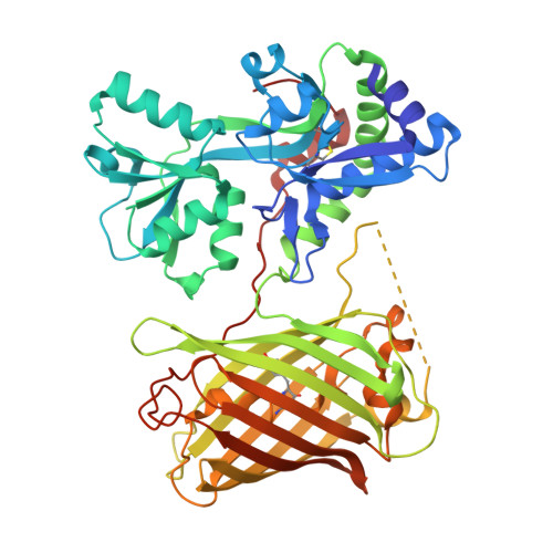 | |
UniProt | |||||
Entity Groups | |||||
| Sequence Clusters | 30% Identity50% Identity70% Identity90% Identity95% Identity100% Identity | ||||
| UniProt Group | A0A0H2UXX1 | ||||
Sequence AnnotationsExpand | |||||
Reference Sequence | |||||
| Ligands 1 Unique | |||||
|---|---|---|---|---|---|
| ID | Chains | Name / Formula / InChI Key | 2D Diagram | 3D Interactions | |
| GLU (Subject of Investigation/LOI) Download:Ideal Coordinates CCD File | B [auth A] | GLUTAMIC ACID C5 H9 N O4 WHUUTDBJXJRKMK-VKHMYHEASA-N |  | ||
| Modified Residues 1 Unique | |||||
|---|---|---|---|---|---|
| ID | Chains | Type | Formula | 2D Diagram | Parent |
| GYS Query on GYS | A | L-PEPTIDE LINKING | C14 H15 N3 O5 |  | SER, TYR, GLY |
| Length ( Å ) | Angle ( ˚ ) |
|---|---|
| a = 68.76 | α = 90 |
| b = 100.84 | β = 90 |
| c = 73.29 | γ = 90 |
| Software Name | Purpose |
|---|---|
| PHENIX | refinement |
| XDS | data reduction |
| XSCALE | data scaling |
| PHASER | phasing |
| Funding Organization | Location | Grant Number |
|---|---|---|
| Max Planck Society | Germany | -- |


