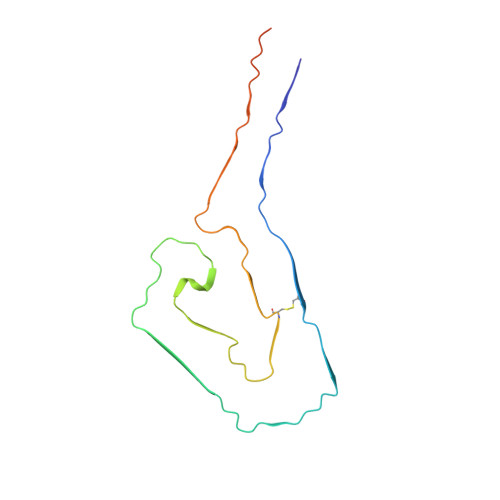
Helical superstructures between amyloid and collagen in cardiac fibrils from a patient with AL amyloidosis.
Schulte, T., Chaves-Sanjuan, A., Speranzini, V., Sicking, K., Milazzo, M., Mazzini, G., Rognoni, P., Caminito, S., Milani, P., Marabelli, C., Corbelli, A., Diomede, L., Fiordaliso, F., Anastasia, L., Pappone, C., Merlini, G., Bolognesi, M., Nuvolone, M., Fernandez-Busnadiego, R., Palladini, G., Ricagno, S.(2024) Nat Commun 15: 6359-6359
- PubMed: 39069558
- DOI: https://doi.org/10.1038/s41467-024-50686-2
- Primary Citation of Related Structures:
9FAA, 9FAC - PubMed Abstract:
Systemic light chain (LC) amyloidosis (AL) is a disease where organs are damaged by an overload of a misfolded patient-specific antibody-derived LC, secreted by an abnormal B cell clone. The high LC concentration in the blood leads to amyloid deposition at organ sites. Indeed, cryogenic electron microscopy (cryo-EM) has revealed unique amyloid folds for heart-derived fibrils taken from different patients. Here, we present the cryo-EM structure of heart-derived AL amyloid (AL59) from another patient with severe cardiac involvement. The double-layered structure displays a u-shaped core that is closed by a β-arc lid and extended by a straight tail. Noteworthy, the fibril harbours an extended constant domain fragment, thus ruling out the variable domain as sole amyloid building block. Surprisingly, the fibrils were abundantly concatenated with a proteinaceous polymer, here identified as collagen VI (COLVI) by immuno-electron microscopy (IEM) and mass-spectrometry. Cryogenic electron tomography (cryo-ET) showed how COLVI wraps around the amyloid forming a helical superstructure, likely stabilizing and protecting the fibrils from clearance. Thus, here we report structural evidence of interactions between amyloid and collagen, potentially signifying a distinct pathophysiological mechanism of amyloid deposits.
- Institute of Molecular and Translational Cardiology, IRCCS Policlinico San Donato, Piazza Malan 2, 20097, San Donato Milanese, Italy.
Organizational Affiliation:


