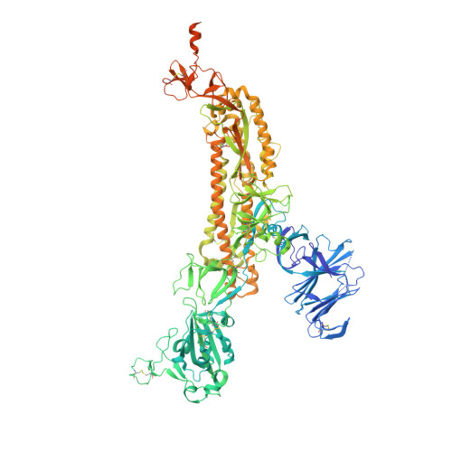
BA.2.87.1 represents a major shift in the BA.2 lineage of severe acute respiratory syndrome coronavirus 2 (SARS-CoV-2) and is unusual in having two lengthy deletions of polypeptide in the spike (S) protein, one of which removes a beta-strand. Here we investigate its neutralization by a variety of sera from infected and vaccinated individuals and determine its spike (S) ectodomain structure. The BA.2.87.1 receptor binding domain (RBD) is structurally conserved and the RBDs are tightly packed in an "all-down" conformation with a small rotation relative to the trimer axis as compared to the closest previously observed conformation. The N-terminal domain (NTD) maintains a remarkably similar structure overall; however, the rearrangements resulting from the deletions essentially destroy the so-called supersite epitope and eliminate one glycan site, while a mutation creates an additional glycan site, effectively shielding another NTD epitope. BA.2.87.1 is relatively easily neutralized but acquisition of additional mutations in the RBD could increase antibody escape allowing it to become a dominant sub-lineage.
Organizational Affiliation:
Division of Structural Biology, Nuffield Department of Medicine, University of Oxford, The Centre for Human Genetics, Oxford, UK.
Chinese Academy of Medical Science (CAMS) Oxford Institute (COI), University of Oxford, Oxford, UK; Centre for Human Genetics, Nuffield Department of Medicine, University of Oxford, Oxford, UK.
NDM Centre For Global Health Research, Nuffield Department of Medicine, University of Oxford, Oxford, UK; Peter Medawar Building for Pathogen Research, University of Oxford, Oxford, UK.
Oxford University Hospitals NHS Foundation Trust, Oxford, UK; Radcliffe Department of Medicine, University of Oxford, Oxford, UK.
Oxford University Hospitals NHS Foundation Trust, Oxford, UK.
Peter Medawar Building for Pathogen Research, University of Oxford, Oxford, UK; NIHR Oxford Biomedical Research Centre, Oxford University Hospitals NHS Foundation Trust, Oxford, UK; Translational Gastroenterology Unit, Nuffield Department of Medicine, University of Oxford, Oxford, UK.
Peter Medawar Building for Pathogen Research, University of Oxford, Oxford, UK; NIHR Oxford Biomedical Research Centre, Oxford University Hospitals NHS Foundation Trust, Oxford, UK; Translational Gastroenterology Unit, Nuffield Department of Medicine, University of Oxford, Oxford, UK; Mahidol-Oxford Tropical Medicine Research Unit, Bangkok, Thailand.
Chinese Academy of Medical Science (CAMS) Oxford Institute (COI), University of Oxford, Oxford, UK; Centre for Human Genetics, Nuffield Department of Medicine, University of Oxford, Oxford, UK; Mahidol-Oxford Tropical Medicine Research Unit, Bangkok, Thailand. Electronic address: juthathip.mongkolsapaya@well.ox.ac.uk.
Division of Structural Biology, Nuffield Department of Medicine, University of Oxford, The Centre for Human Genetics, Oxford, UK. Electronic address: liz@strubi.ox.ac.uk.
Division of Structural Biology, Nuffield Department of Medicine, University of Oxford, The Centre for Human Genetics, Oxford, UK. Electronic address: ren@strubi.ox.ac.uk.
Division of Structural Biology, Nuffield Department of Medicine, University of Oxford, The Centre for Human Genetics, Oxford, UK; Chinese Academy of Medical Science (CAMS) Oxford Institute (COI), University of Oxford, Oxford, UK; Diamond Light Source Ltd, Harwell Science & Innovation Campus, Didcot, UK. Electronic address: dave@strubi.ox.ac.uk.
Chinese Academy of Medical Science (CAMS) Oxford Institute (COI), University of Oxford, Oxford, UK; Centre for Human Genetics, Nuffield Department of Medicine, University of Oxford, Oxford, UK. Electronic address: gavin.screaton@medsci.ox.ac.uk.
AA [auth B] BA [auth B] CA [auth B] DA [auth B] EA [auth B]
AA [auth B], BA [auth B], CA [auth B], DA [auth B], EA [auth B], FA [auth B], GA [auth B], HA [auth B], IA [auth B], JA [auth B], KA [auth B], LA [auth B], M [auth A], MA [auth C], N [auth A], NA [auth C], O [auth A], OA [auth C], P [auth A], PA [auth C], Q [auth A], QA [auth C], R [auth A], RA [auth C], S [auth A], SA [auth C], T [auth A], TA [auth C], U [auth A], UA [auth C], V [auth A], VA [auth C], W [auth A], WA [auth C], X [auth A], XA [auth C], Y [auth A], YA [auth C], Z [auth B]
2-acetamido-2-deoxy-beta-D-glucopyranose C8 H15 N O6 OVRNDRQMDRJTHS-FMDGEEDCSA-N