9F8X

Low-dose structure of Marinobacter nauticus nitrous oxide reductase

  • Classification: OXIDOREDUCTASE
  • Organism(s): Marinobacter nauticus
  • Mutation(s): Yes 

  • Deposited: 2024-05-07 Released: 2024-06-19 
  • Deposition Author(s): Einsle, O., Pomowski, A.
  • Funding Organization(s): German Research Foundation (DFG), European Research Council (ERC), Foundation for Science and Technology (FCT), European Molecular Biology Organization (EMBO)

Experimental Data Snapshot

  • Method: X-RAY DIFFRACTION
  • Resolution: 1.50 Å
  • R-Value Free: 0.152 
  • R-Value Work: 0.130 

Starting Model: experimental
View more details

wwPDB Validation   3D Report Full Report


This is version 1.0 of the entry. See complete history


Literature

Revisiting the metal sites of nitrous oxide reductase in a low-dose structure from Marinobacter nauticus.

Pomowski, A.Dell'Acqua, S.Wust, A.Pauleta, S.R.Moura, I.Einsle, O.

(2024) J Biol Inorg Chem 29: 279-290

  • DOI: https://doi.org/10.1007/s00775-024-02056-y
  • Primary Citation of Related Structures:  
    9F8X

  • PubMed Abstract: 

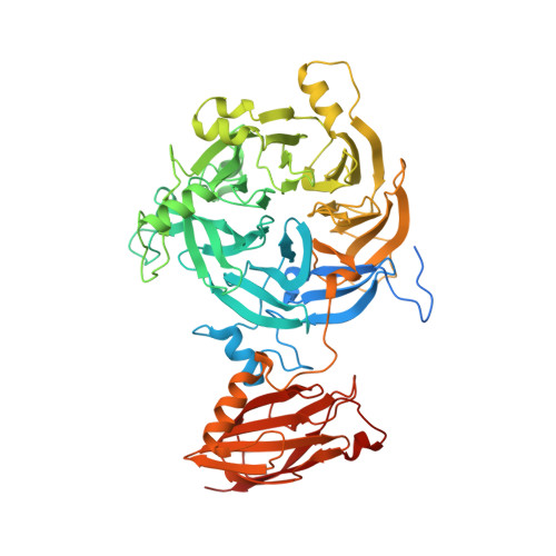
    Copper-containing nitrous oxide reductase catalyzes a 2-electron reduction of the green-house gas N 2 O to yield N 2 . It contains two metal centers, the binuclear electron transfer site Cu A , and the unique, tetranuclear Cu Z center that is the site of substrate binding. Different forms of the enzyme were described previously, representing variations in oxidation state and composition of the metal sites. Hypothesizing that many reported discrepancies in the structural data may be due to radiation damage during data collection, we determined the structure of anoxically isolated Marinobacter nauticus N 2 OR from diffraction data obtained with low-intensity X-rays from an in-house rotating anode generator and an image plate detector. The data set was of exceptional quality and yielded a structure at 1.5 Å resolution in a new crystal form. The Cu A site of the enzyme shows two distinct conformations with potential relevance for intramolecular electron transfer, and the Cu Z cluster is present in a [4Cu:2S] configuration. In addition, the structure contains three additional types of ions, and an analysis of anomalous scattering contributions confirms them to be Ca 2+ , K + , and Cl - . The uniformity of the present structure supports the hypothesis that many earlier analyses showed inhomogeneities due to radiation effects. Adding to the earlier description of the same enzyme with a [4Cu:S] Cu Z site, a mechanistic model is presented, with a structurally flexible Cu Z center that does not require the complete dissociation of a sulfide prior to N 2 O binding.


  • Organizational Affiliation

    Institute for Biochemistry, Albert-Ludwigs-University Freiburg, Albertstrasse 21, 79104, Freiburg, Germany.


Macromolecules
Find similar proteins by:  (by identity cutoff)  |  3D Structure
Entity ID: 1
MoleculeChains Sequence LengthOrganismDetailsImage
Nitrous-oxide reductase
A, B, C, D
631Marinobacter nauticusMutation(s): 3 
EC: 1.7.2.4
UniProt
Find proteins for A0A368XMX8 (Marinobacter nauticus)
Explore A0A368XMX8 
Go to UniProtKB:  A0A368XMX8
Entity Groups  
Sequence Clusters30% Identity50% Identity70% Identity90% Identity95% Identity100% Identity
UniProt GroupA0A368XMX8
Sequence Annotations
Expand
  • Reference Sequence
Small Molecules
Ligands 6 Unique
IDChains Name / Formula / InChI Key2D Diagram3D Interactions
CUK (Subject of Investigation/LOI)
Query on CUK

Download Ideal Coordinates CCD File 
AA [auth C],
F [auth A],
IA [auth D],
M [auth B]
[4Cu:2S] cluster
Cu4 S2
PDBAWKBNXYUXTL-UHFFFAOYSA-N
CUA
Query on CUA

Download Ideal Coordinates CCD File 
E [auth A],
HA [auth D],
L [auth B],
Z [auth C]
DINUCLEAR COPPER ION
Cu2
ALKZAGKDWUSJED-UHFFFAOYSA-N
EDO
Query on EDO

Download Ideal Coordinates CCD File 
EA [auth C]
FA [auth C]
GA [auth C]
J [auth A]
K [auth A]
EA [auth C],
FA [auth C],
GA [auth C],
J [auth A],
K [auth A],
MA [auth D],
NA [auth D],
T [auth B],
U [auth B],
V [auth B],
W [auth B],
X [auth B],
Y [auth B]
1,2-ETHANEDIOL
C2 H6 O2
LYCAIKOWRPUZTN-UHFFFAOYSA-N
CA
Query on CA

Download Ideal Coordinates CCD File 
BA [auth C]
DA [auth C]
G [auth A]
I [auth A]
JA [auth D]
BA [auth C],
DA [auth C],
G [auth A],
I [auth A],
JA [auth D],
LA [auth D],
N [auth B],
P [auth B]
CALCIUM ION
Ca
BHPQYMZQTOCNFJ-UHFFFAOYSA-N
CL
Query on CL

Download Ideal Coordinates CCD File 
CA [auth C],
H [auth A],
KA [auth D],
O [auth B]
CHLORIDE ION
Cl
VEXZGXHMUGYJMC-UHFFFAOYSA-M
NA
Query on NA

Download Ideal Coordinates CCD File 
Q [auth B],
R [auth B],
S [auth B]
SODIUM ION
Na
FKNQFGJONOIPTF-UHFFFAOYSA-N
Experimental Data & Validation

Experimental Data

  • Method: X-RAY DIFFRACTION
  • Resolution: 1.50 Å
  • R-Value Free: 0.152 
  • R-Value Work: 0.130 
  • Space Group: P 1
Unit Cell:
Length ( Å )Angle ( ˚ )
a = 65.26α = 81.384
b = 69.375β = 77.918
c = 153.188γ = 88.608
Software Package:
Software NamePurpose
REFMACrefinement
DENZOdata reduction
Aimlessdata scaling
MOLREPphasing

Structure Validation

View Full Validation Report


Entry History & Funding Information

Deposition Data


Funding OrganizationLocationGrant Number
German Research Foundation (DFG)Germany235777276
German Research Foundation (DFG)Germany311061829
European Research Council (ERC)European Union310656
Foundation for Science and Technology (FCT)PortugalUIDP/04378/2020
Foundation for Science and Technology (FCT)PortugalUIDB/04378/2020
Foundation for Science and Technology (FCT)PortugalLA/P/0140/2020
Foundation for Science and Technology (FCT)PortugalUIDB/50006/2020
Foundation for Science and Technology (FCT)PortugalUIDP/50006/2020
Foundation for Science and Technology (FCT)Portugal2022.01152.PTDC
European Molecular Biology Organization (EMBO)European UnionASTF 282.00-2010

Revision History  (Full details and data files)

  • Version 1.0: 2024-06-19
    Type: Initial release