9F1I

Crystal structure of a first-in-class antibody for alpha-1,6-fucosylated prostate-specific antigen, target bound


Experimental Data Snapshot

  • Method: X-RAY DIFFRACTION
  • Resolution: 1.38 Å
  • R-Value Free: 0.186 
  • R-Value Work: 0.155 
  • R-Value Observed: 0.156 

Starting Model: experimental
View more details

wwPDB Validation   3D Report Full Report


Ligand Structure Quality Assessment 


This is version 1.0 of the entry. See complete history


Literature

Development of a first-in-class antibody and a specific assay for alpha-1,6-fucosylated prostate-specific antigen.

Halldorsson, S.Hillringhaus, L.Hojer, C.Muranyi, A.Schraeml, M.Lange, M.S.Tabares, G.

(2024) Sci Rep 14: 16512-16512

  • DOI: https://doi.org/10.1038/s41598-024-67545-1
  • Primary Citation of Related Structures:  
    9F18, 9F1I

  • PubMed Abstract: 

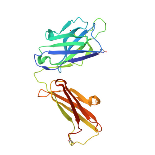
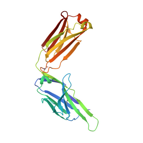
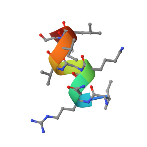
    Prostate-specific antigen (PSA) levels are widely used to screen for prostate cancer, yet the test has poor sensitivity, specificity and predictive value, which leads to overdiagnosis and overtreatment. Alterations in the glycosylation status of PSA, including fucosylation, may offer scope for an improved biomarker. We sought to generate a monoclonal antibody (mAb) targeting α-1,6-fucosylated PSA (fuc-PSA) and to develop a tissue-based immunological assay for fuc-PSA detection. Immunogens representing fuc-PSA were used for immunisation and resultant mAbs were extensively characterised. The mAbs reacted specifically with fuc-PSA-specific glycopeptide, but not with aglycosylated PSA or glycan without the PSA peptide. Reactivity was confirmed using high-throughput surface plasmon resonance spectroscopy. X-ray crystallography investigations showed that the mAbs bound to an α-helical form of the peptide, whereas the native PSA epitope is linear. Protein unfolding was required for detection of fuc-PSA in patient samples. Peptide inhibition of fuc-PSA mAbs was observed with positive screening reagents, and target epitope specificity was observed in formalin-fixed, paraffin-embedded tissue samples. This research introduces a well-characterised, first-in-class antibody targeting fuc-PSA and presents the first crystal structure of an antibody demonstrating glycosylation-specific binding to a peptide.


  • Organizational Affiliation

    Pharma Research and Early Development, F. Hoffmann-La Roche AG, Basel, Switzerland.


Macromolecules
Find similar proteins by:  (by identity cutoff)  |  3D Structure
Entity ID: 1
MoleculeChains Sequence LengthOrganismDetailsImage
Heavy chain rabbit fabA [auth H],
B [auth I]
230Oryctolagus cuniculusMutation(s): 0 
Entity Groups  
Sequence Clusters30% Identity50% Identity70% Identity90% Identity95% Identity100% Identity
Sequence Annotations
Expand
  • Reference Sequence
Find similar proteins by:  (by identity cutoff)  |  3D Structure
Entity ID: 2
MoleculeChains Sequence LengthOrganismDetailsImage
Light chain rabbit fabC [auth L],
D [auth M]
217Oryctolagus cuniculusMutation(s): 0 
Entity Groups  
Sequence Clusters30% Identity50% Identity70% Identity90% Identity95% Identity100% Identity
Sequence Annotations
Expand
  • Reference Sequence

Find similar proteins by:  Sequence   |   3D Structure  

Entity ID: 3
MoleculeChains Sequence LengthOrganismDetailsImage
Prostate specific antigenE [auth P],
F [auth Q]
10Homo sapiensMutation(s): 0 
UniProt
Find proteins for Q8NCW4 (Homo sapiens)
Explore Q8NCW4 
Go to UniProtKB:  Q8NCW4
Entity Groups  
Sequence Clusters30% Identity50% Identity70% Identity90% Identity95% Identity100% Identity
UniProt GroupQ8NCW4
Glycosylation
Glycosylation Sites: 1
Sequence Annotations
Expand
  • Reference Sequence
Oligosaccharides

Help

Entity ID: 4
MoleculeChains Length2D Diagram Glycosylation3D Interactions
alpha-L-fucopyranose-(1-6)-2-acetamido-2-deoxy-beta-D-glucopyranoseG [auth A],
H [auth C]
2N-Glycosylation
Glycosylation Resources
GlyTouCan:  G86851RC
GlyCosmos:  G86851RC
GlyGen:  G86851RC
Experimental Data & Validation

Experimental Data

  • Method: X-RAY DIFFRACTION
  • Resolution: 1.38 Å
  • R-Value Free: 0.186 
  • R-Value Work: 0.155 
  • R-Value Observed: 0.156 
  • Space Group: P 21 21 21
Unit Cell:
Length ( Å )Angle ( ˚ )
a = 72.31α = 90
b = 87.395β = 90
c = 150.427γ = 90
Software Package:
Software NamePurpose
PHENIXrefinement
XDSdata reduction
XSCALEdata scaling
PHASERphasing

Structure Validation

View Full Validation Report


Ligand Structure Quality Assessment 


Entry History & Funding Information

Deposition Data


Funding OrganizationLocationGrant Number
F. Hoffmann-La Roche LTDSwitzerland--

Revision History  (Full details and data files)

  • Version 1.0: 2024-07-31
    Type: Initial release