9ETO | pdb_00009eto

PsiK from Psilocybe cubensis


Experimental Data Snapshot

  • Method: X-RAY DIFFRACTION
  • Resolution: 2.54 Å
  • R-Value Free: 
    0.243 (Depositor), 0.245 (DCC) 
  • R-Value Work: 
    0.197 (Depositor), 0.204 (DCC) 

Starting Model: in silico
View more details

wwPDB Validation 3D Report Full Report

Validation slider image for 9ETO

Ligand Structure Quality Assessment 


This is version 1.1 of the entry. See complete history

Literature

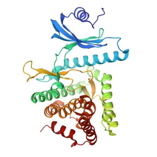
Substrate recognition by the 4-hydroxytryptamine kinase PsiK in psilocybin biosynthesis.

Rogge, K.Wagner, T.J.Hoffmeister, D.Rupp, B.Werten, S.

(2025) FEBS Lett 599: 447-455

  • DOI: https://doi.org/10.1002/1873-3468.15042
  • Primary Citation Related Structures: 
    9ETO

  • PubMed Abstract: 

    Psilocybin, the natural hallucinogen from Psilocybe (magic) mushrooms, is a highly promising drug candidate for the treatment of depression and several other mental health conditions. Biosynthesis of psilocybin from the amino acid l-tryptophan involves four strictly sequential modifications. The third of these, ATP-dependent phosphorylation of the intermediate 4-hydroxytryptamine, is catalysed by PsiK. Here we present a crystallographic analysis and a structure-based mutagenesis study of this kinase, providing insight into its mode of substrate recognition. The results of our work will support future bioengineering efforts aimed at generating variants of psilocybin with enhanced therapeutic properties.


  • Organizational Affiliation
    • Institute of Pharmacy, Friedrich Schiller University, Jena, Germany.

Macromolecule Content 

  • Total Structure Weight: 83.28 kDa 
  • Atom Count: 5,797 
  • Modeled Residue Count: 726 
  • Deposited Residue Count: 728 
  • Unique protein chains: 1

Macromolecules

Find similar proteins by:|  3D Structure
Entity ID: 1
MoleculeChains  Sequence LengthOrganismDetailsImage
4-hydroxytryptamine kinase
A, B
364Psilocybe cubensisMutation(s): 0 
Gene Names: psiK
EC: 2.7.1 (PDB Primary Data), 2.7.1.222 (PDB Primary Data)
UniProt
Find proteins for P0DPA8 (Psilocybe cubensis)
Explore P0DPA8 
Go to UniProtKB:  P0DPA8
Entity Groups
Sequence Clusters30% Identity50% Identity70% Identity90% Identity95% Identity100% Identity
UniProt GroupP0DPA8
Sequence Annotations
Expand
Reference Sequence

Small Molecules

Ligands 4 Unique
IDChains Name / Formula / InChI Key2D Diagram3D Interactions
RWB

Query on RWB


Download:Ideal Coordinates CCD File
L [auth A]dodecaethylene glycol monomethyl ether
C25 H52 O13
PLQZJIIDLZRWBG-UHFFFAOYSA-N
PEG
(Subject of Investigation/LOI)

Query on PEG


Download:Ideal Coordinates CCD File
C [auth A],
M [auth B]
DI(HYDROXYETHYL)ETHER
C4 H10 O3
MTHSVFCYNBDYFN-UHFFFAOYSA-N
SO4

Query on SO4


Download:Ideal Coordinates CCD File
D [auth A]
E [auth A]
F [auth A]
G [auth A]
K [auth A]
D [auth A],
E [auth A],
F [auth A],
G [auth A],
K [auth A],
N [auth B],
O [auth B],
P [auth B],
Q [auth B],
R [auth B]
SULFATE ION
O4 S
QAOWNCQODCNURD-UHFFFAOYSA-L
CL

Query on CL


Download:Ideal Coordinates CCD File
H [auth A],
I [auth A],
J [auth A],
S [auth B],
T [auth B]
CHLORIDE ION
Cl
VEXZGXHMUGYJMC-UHFFFAOYSA-M

Experimental Data & Validation

Experimental Data

  • Method: X-RAY DIFFRACTION
  • Resolution: 2.54 Å
  • R-Value Free:  0.243 (Depositor), 0.245 (DCC) 
  • R-Value Work:  0.197 (Depositor), 0.204 (DCC) 
Space Group: C 1 2 1
Unit Cell:
Length ( Å )Angle ( ˚ )
a = 111.82α = 90
b = 70.14β = 95.837
c = 118.9γ = 90
Software Package:
Software NamePurpose
REFMACrefinement
XDSdata reduction
XSCALEdata scaling
PHASERphasing

Structure Validation

View Full Validation Report


Ligand Structure Quality Assessment 


Entry History 

& Funding Information

Deposition Data


Funding OrganizationLocationGrant Number
Austrian Science FundAustriaI-5192

Revision History  (Full details and data files)

  • Version 1.0: 2024-11-06
    Type: Initial release
  • Version 1.1: 2025-02-19
    Changes: Database references