Structures of alpha-galactosaminidases from the CAZy GH114 family and homologs defining a new GH191 family of glycosidases.
Roth, C., Moroz, O.V., Miranda, S.A.D., Jahn, L., Blagova, E.V., Lebedev, A.A., Segura, D.R., Stringer, M.A., Friis, E.P., Franco Cairo, J.P.L., Davies, G.J., Wilson, K.S.(2025) Acta Crystallogr D Struct Biol 81: 234-251
- PubMed: 40232846 Search on PubMed
- DOI: https://doi.org/10.1107/S2059798325002864
- Primary Citation Related Structures:
9EP5, 9EP6, 9EUX, 9EUZ - PubMed Abstract:
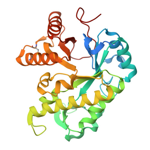
Endo-galactosaminidases are an underexplored family of enzymes involved in the degradation of galactosaminogalactan (GAG) and other galactosamine-containing cationic exopolysaccharides produced by fungi and bacteria. These exopolysaccharides are part of the cell wall and extracellular matrix of microbial communities. Currently, these galactosaminidases are found in three distinct CAZy families: GH114, GH135 and GH166. Despite the widespread occurrence of these enzymes in nearly all bacterial and fungal clades, only limited biochemical and structural data are available for these three groups. To expand our knowledge of endo-galactosaminidases, we selected several sequences predicted to encode endo-galactosaminidases and produced them recombinantly for structural and functional studies. Only very few predicted proteins could be produced in soluble form, and activity against bacterial Pel (pellicle) polysaccharide could only be confirmed for one enzyme. Here, we report the structures of two bacterial and one fungal enzyme. Whereas the fungal enzyme belongs to family GH114, the two bacterial enzymes do not lie in the current GH families but instead define a new family, GH191. During structure solution we realized that crystals of all three enzymes had various defects including twinning and partial disorder, which in the case of a more severe pathology in one of the structures required the design of a specialized refinement/model-building protocol. Comparison of the structures revealed several features that might be responsible for the described activity pattern and substrate specificity compared with other GAG-degrading enzymes.
- Department for Biomolecular Systems, Carbohydrates Structure and Function, Max Planck Institute of Colloids and Interfaces, Arnimallee 22, 14195 Berlin, Germany.
Organizational Affiliation: