9EOV | pdb_00009eov

Crystal structure of domains I and II from the outer membrane cytochrome MtrC


Experimental Data Snapshot

  • Method: X-RAY DIFFRACTION
  • Resolution: 1.80 Å
  • R-Value Free: 
    0.221 (Depositor), 0.221 (DCC) 
  • R-Value Work: 
    0.188 (Depositor), 0.188 (DCC) 
  • R-Value Observed: 
    0.189 (Depositor) 

Starting Model: experimental
View more details

wwPDB Validation 3D Report Full Report

Validation slider image for 9EOV

Ligand Structure Quality Assessment 


This is version 1.0 of the entry. See complete history

Literature

Extracellular catalysis of environmental substrates by Shewanella oneidensis MR-1 occurs via active sites on the C-terminal domains of MtrC.

Morales-Florez, A.Lockwood, C.W.J.Nash, B.W.Edwards, M.J.van Wonderen, J.H.Sachdeva, A.Butt, J.N.Clarke, T.A.

(2025) Protein Sci 34: e70243-e70243

  • DOI: https://doi.org/10.1002/pro.70243
  • Primary Citation Related Structures: 
    9EOV

  • PubMed Abstract: 

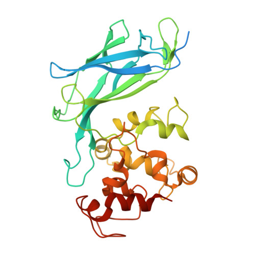
    The Gram-negative Shewanellaceae family is well known for its ability to transfer catabolically derived electrons to extracellular terminal electron acceptors through electron conduits that permeate the outer membrane. The primary conduit is MtrCAB, a trimeric porin-cytochrome complex that contains the cell surface exposed decaheme cytochrome MtrC. This donates electrons to extracellular substrates, including OmcA, soluble metals, organic electron shuttles, and insoluble metal oxides. However, it is not clear whether this broad substrate specificity requires specific sites for binding and reduction, or whether reduction occurs through non-specific interactions near exposed hemes on the cytochrome surface. Shewanella oneidensis MtrC is composed of four domains, with the hemes closely packed and distributed evenly between domains II and IV. The domains are arranged to allow electron transport across the cytochrome via interdomain electron transfer, but the significance of this conserved feature is not understood. Here we use site-directed mutagenesis to generate an MtrC variant that is comprised only of domains I and II (MtrC DI,II ). The properties of this MtrC DI,II are effectively identical to domains I and II of full-length MtrC. Whole-cell assays revealed that S. oneidensis cells replacing full-length MtrC with MtrC DI,II had significantly lower rates of OmcA, flavin mononucleotide, and Fe(III) citrate reduction. Our results demonstrate that MtrC domains III and IV contain sites for association of specific substrates, enabling the reduction of extracellular electron acceptors in S. oneidensis.


  • Organizational Affiliation
    • School of Biological Sciences, University of East Anglia, Norwich, UK.

Macromolecule Content 

  • Total Structure Weight: 37.82 kDa 
  • Atom Count: 2,723 
  • Modeled Residue Count: 278 
  • Deposited Residue Count: 323 
  • Unique protein chains: 1

Macromolecules

Find similar proteins by:|  3D Structure
Entity ID: 1
MoleculeChains  Sequence LengthOrganismDetailsImage
Extracellular iron oxide respiratory system surface decaheme cytochrome c component MtrC323Shewanella oneidensis MR-1Mutation(s): 0 
Gene Names: mtrCSO_1778
UniProt
Find proteins for Q8EG34 (Shewanella oneidensis (strain ATCC 700550 / JCM 31522 / CIP 106686 / LMG 19005 / NCIMB 14063 / MR-1))
Explore Q8EG34 
Go to UniProtKB:  Q8EG34
Entity Groups
Sequence Clusters30% Identity50% Identity70% Identity90% Identity95% Identity100% Identity
UniProt GroupQ8EG34
Sequence Annotations
Expand
Reference Sequence

Small Molecules

Ligands 3 Unique
IDChains Name / Formula / InChI Key2D Diagram3D Interactions
HEC
(Subject of Investigation/LOI)

Query on HEC


Download:Ideal Coordinates CCD File
B [auth A],
C [auth A],
D [auth A],
E [auth A],
F [auth A]
HEME C
C34 H34 Fe N4 O4
HXQIYSLZKNYNMH-LJNAALQVSA-N
EDO

Query on EDO


Download:Ideal Coordinates CCD File
H [auth A],
I [auth A],
J [auth A]
1,2-ETHANEDIOL
C2 H6 O2
LYCAIKOWRPUZTN-UHFFFAOYSA-N
MG

Query on MG


Download:Ideal Coordinates CCD File
G [auth A]MAGNESIUM ION
Mg
JLVVSXFLKOJNIY-UHFFFAOYSA-N

Experimental Data & Validation

Experimental Data

  • Method: X-RAY DIFFRACTION
  • Resolution: 1.80 Å
  • R-Value Free:  0.221 (Depositor), 0.221 (DCC) 
  • R-Value Work:  0.188 (Depositor), 0.188 (DCC) 
  • R-Value Observed: 0.189 (Depositor) 
Space Group: P 21 21 21
Unit Cell:
Length ( Å )Angle ( ˚ )
a = 74.275α = 90
b = 77.35β = 90
c = 96.531γ = 90
Software Package:
Software NamePurpose
PHENIXrefinement
Aimlessdata scaling
xia2data reduction
PHASERphasing

Structure Validation

View Full Validation Report


Ligand Structure Quality Assessment 


Entry History 

& Funding Information

Deposition Data


Funding OrganizationLocationGrant Number
Biotechnology and Biological Sciences Research Council (BBSRC)United KingdomBB/T008717/1

Revision History  (Full details and data files)

  • Version 1.0: 2025-08-13
    Type: Initial release