9ENT

SSX structure of Autotaxin in cryogenic conditions


Experimental Data Snapshot

  • Method: X-RAY DIFFRACTION
  • Resolution: 2.20 Å
  • R-Value Free: 0.236 
  • R-Value Work: 0.194 
  • R-Value Observed: 0.196 

Starting Model: experimental
View more details

wwPDB Validation   3D Report Full Report


Ligand Structure Quality Assessment 


This is version 1.0 of the entry. See complete history


Literature

In situ serial crystallography facilitates 96-well plate structural analysis at low symmetry.

Foos, N.Florial, J.B.Eymery, M.Sinoir, J.Felisaz, F.Oscarsson, M.Beteva, A.Bowler, M.W.Nurizzo, D.Papp, G.Soler-Lopez, M.Nanao, M.Basu, S.McCarthy, A.A.

(2024) IUCrJ 

  • DOI: https://doi.org/10.1107/S2052252524005785
  • Primary Citation of Related Structures:  
    9ENT, 9EU5

  • PubMed Abstract: 

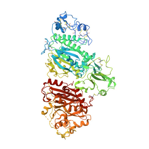
    The advent of serial crystallography has rejuvenated and popularized room-temperature X-ray crystal structure determination. Structures determined at physiological temperature reveal protein flexibility and dynamics. In addition, challenging samples (e.g. large complexes, membrane proteins and viruses) form fragile crystals that are often difficult to harvest for cryo-crystallography. Moreover, a typical serial crystallography experiment requires a large number of microcrystals, mainly achievable through batch crystallization. Many medically relevant samples are expressed in mammalian cell lines, producing a meager quantity of protein that is incompatible with batch crystallization. This can limit the scope of serial crystallography approaches. Direct in situ data collection from a 96-well crystallization plate enables not only the identification of the best diffracting crystallization condition but also the possibility for structure determination under ambient conditions. Here, we describe an in situ serial crystallography (iSX) approach, facilitating direct measurement from crystallization plates mounted on a rapidly exchangeable universal plate holder deployed at a microfocus beamline, ID23-2, at the European Synchrotron Radiation Facility. We applied our iSX approach on a challenging project, autotaxin, a therapeutic target expressed in a stable human cell line, to determine the structure in the lowest-symmetry P1 space group at 3.0 Å resolution. Our in situ data collection strategy provided a complete dataset for structure determination while screening various crystallization conditions. Our data analysis reveals that the iSX approach is highly efficient at a microfocus beamline, improving throughput and demonstrating how crystallization plates can be routinely used as an alternative method of presenting samples for serial crystallography experiments at synchrotrons.


  • Organizational Affiliation

    European Molecular Biology Laboratory, Grenoble Outstation, 71 Avenue des Martyrs, 38042 Grenoble, France.


Macromolecules
Find similar proteins by:  (by identity cutoff)  |  3D Structure
Entity ID: 1
MoleculeChains Sequence LengthOrganismDetailsImage
Ectonucleotide pyrophosphatase/phosphodiesterase family member 2862Rattus norvegicusMutation(s): 0 
Gene Names: Enpp2AtxNpps2
EC: 3.1.4.39
UniProt
Find proteins for Q64610 (Rattus norvegicus)
Explore Q64610 
Go to UniProtKB:  Q64610
Entity Groups  
Sequence Clusters30% Identity50% Identity70% Identity90% Identity95% Identity100% Identity
UniProt GroupQ64610
Glycosylation
Glycosylation Sites: 1Go to GlyGen: Q64610-1
Sequence Annotations
Expand
  • Reference Sequence
Oligosaccharides

Help

Entity ID: 2
MoleculeChains Length2D Diagram Glycosylation3D Interactions
2-acetamido-2-deoxy-beta-D-glucopyranose-(1-4)-2-acetamido-2-deoxy-beta-D-glucopyranose-(1-4)-2-acetamido-2-deoxy-beta-D-glucopyranose
B
3N-Glycosylation
Glycosylation Resources
GlyTouCan:  G47362BJ
GlyCosmos:  G47362BJ
GlyGen:  G47362BJ
Small Molecules
Ligands 6 Unique
IDChains Name / Formula / InChI Key2D Diagram3D Interactions
5JK (Subject of Investigation/LOI)
Query on 5JK

Download Ideal Coordinates CCD File 
C [auth A]7alpha-hydroxycholesterol
C27 H46 O2
OYXZMSRRJOYLLO-RVOWOUOISA-N
IOD
Query on IOD

Download Ideal Coordinates CCD File 
H [auth A]
I [auth A]
J [auth A]
K [auth A]
L [auth A]
H [auth A],
I [auth A],
J [auth A],
K [auth A],
L [auth A],
M [auth A],
N [auth A],
O [auth A],
P [auth A],
Q [auth A],
R [auth A],
S [auth A],
T [auth A],
U [auth A],
V [auth A]
IODIDE ION
I
XMBWDFGMSWQBCA-UHFFFAOYSA-M
ZN
Query on ZN

Download Ideal Coordinates CCD File 
E [auth A],
F [auth A]
ZINC ION
Zn
PTFCDOFLOPIGGS-UHFFFAOYSA-N
SCN
Query on SCN

Download Ideal Coordinates CCD File 
D [auth A]THIOCYANATE ION
C N S
ZMZDMBWJUHKJPS-UHFFFAOYSA-M
CA
Query on CA

Download Ideal Coordinates CCD File 
G [auth A]CALCIUM ION
Ca
BHPQYMZQTOCNFJ-UHFFFAOYSA-N
NA
Query on NA

Download Ideal Coordinates CCD File 
W [auth A],
X [auth A],
Y [auth A]
SODIUM ION
Na
FKNQFGJONOIPTF-UHFFFAOYSA-N
Experimental Data & Validation

Experimental Data

Unit Cell:
Length ( Å )Angle ( ˚ )
a = 53.51α = 102.57
b = 61.19β = 99.13
c = 65.01γ = 93.67
Software Package:
Software NamePurpose
PHENIXrefinement
MxCuBEdata collection
PHENIXphasing
CrystFELdata reduction
CrystFELdata scaling

Structure Validation

View Full Validation Report


Ligand Structure Quality Assessment 


Entry History & Funding Information

Deposition Data


Funding OrganizationLocationGrant Number
European Molecular Biology Organization (EMBO)European Union--

Revision History  (Full details and data files)

  • Version 1.0: 2024-07-31
    Type: Initial release