9ENS | pdb_00009ens

Cleavage product of vitellogenin from the honey bee hemolymph


Experimental Data Snapshot

  • Method: ELECTRON MICROSCOPY
  • Resolution: 3.00 Å
  • Aggregation State: PARTICLE 
  • Reconstruction Method: SINGLE PARTICLE 

wwPDB Validation 3D Report Full Report

Validation slider image for 9ENS

This is version 1.3 of the entry. See complete history

Literature

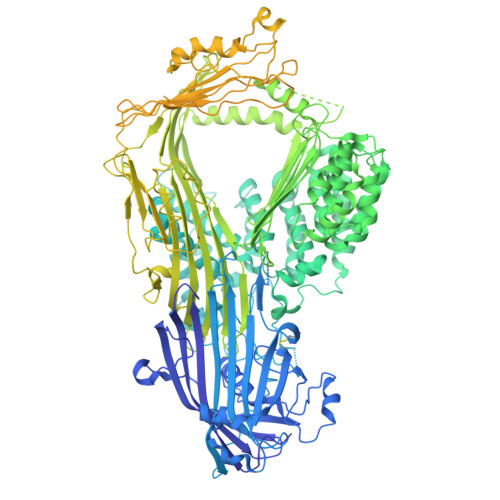
Cryo-EM structure of native honey bee vitellogenin.

Montserrat-Canals, M.Schnelle, K.Leipart, V.Halskau, O.Amdam, G.V.Moeller, A.Cunha, E.S.Luecke, H.

(2025) Nat Commun 16: 5736-5736

  • DOI: https://doi.org/10.1038/s41467-025-58575-y
  • Primary Citation Related Structures: 
    9ENR, 9ENS

  • PubMed Abstract: 

    Vitellogenin (Vg) is the main yolk precursor lipoprotein in almost all egg-laying animals. In addition, along its evolutionary history, Vg has developed a range of new functions in different taxa. In the honey bee, Vg has functions related to immunity, antioxidant protection, social behavior and longevity. However, the molecular mechanisms underlying Vg functionalities are still poorly understood. Here, we report the cryo-EM structure of full-length honey bee Vg, one-step purified directly from hemolymph. The structure provides structural insights into the overall domain architecture, including the lipid binding cavity and the previously uncharacterized von Willebrand factor type D domain. A domain of unknown function has been identified as a C-terminal cystine knot domain based on structural homology. Information about post-translational modifications, cleavage products, metal and lipid binding allow an improved understanding of the mechanisms underlying the range of Vg functionalities. The findings have numerous implications for the structure-function relationship of vitellogenins of other species as well as members of the same protein superfamily, which share the same structural elements.


  • Organizational Affiliation
    • Department of Chemistry, University of Oslo, Oslo, Norway.

Macromolecule Content 

  • Total Structure Weight: 202.22 kDa 
  • Atom Count: 9,076 
  • Modeled Residue Count: 1,161 
  • Deposited Residue Count: 1,770 
  • Unique protein chains: 1

Macromolecules

Find similar proteins by:|  3D Structure
Entity ID: 1
MoleculeChains  Sequence LengthOrganismDetailsImage
Vitellogenin1,770Apis ceranaMutation(s): 0 
UniProt
Find proteins for Q868N5 (Apis mellifera)
Explore Q868N5 
Go to UniProtKB:  Q868N5
Entity Groups
Sequence Clusters30% Identity50% Identity70% Identity90% Identity95% Identity100% Identity
UniProt GroupQ868N5
Glycosylation
Glycosylation Sites: 1
Sequence Annotations
Expand
Reference Sequence

Oligosaccharides

Help  
Entity ID: 2
MoleculeChains Length2D Diagram GlycosylationD Interactions
alpha-D-mannopyranose-(1-3)-[alpha-D-mannopyranose-(1-6)]beta-D-mannopyranose-(1-4)-2-acetamido-2-deoxy-beta-D-glucopyranose-(1-4)-2-acetamido-2-deoxy-beta-D-glucopyranose
B
5N-Glycosylation
Glycosylation Resources
GlyTouCan: G22768VO
GlyCosmos: G22768VO
GlyGen: G22768VO

Experimental Data & Validation

Experimental Data

  • Method: ELECTRON MICROSCOPY
  • Resolution: 3.00 Å
  • Aggregation State: PARTICLE 
  • Reconstruction Method: SINGLE PARTICLE 
EM Software:
TaskSoftware PackageVersion
MODEL REFINEMENTPHENIX
RECONSTRUCTIONcryoSPARC4.2

Structure Validation

View Full Validation Report


Entry History 

& Funding Information

Deposition Data


Funding OrganizationLocationGrant Number
Norwegian Research CouncilNorway262137
Norwegian Research CouncilNorway335244
German Research Foundation (DFG)GermanySFB 944
German Research Foundation (DFG)GermanySFB 1557
German Research Foundation (DFG)GermanyINST190/196-1 FUGG

Revision History  (Full details and data files)

  • Version 1.0: 2025-03-26
    Type: Initial release
  • Version 1.1: 2025-07-02
    Changes: Data collection
  • Version 1.2: 2025-07-09
    Changes: Data collection, Database references
  • Version 1.3: 2025-07-16
    Changes: Data collection, Database references