9EJ8 | pdb_00009ej8

Structure of the Lactate Monooxygenase mutant Y44N, Y152F, H290S, T181A, F184P


Experimental Data Snapshot

  • Method: X-RAY DIFFRACTION
  • Resolution: 1.42 Å
  • R-Value Free: 
    0.178 (Depositor), 0.176 (DCC) 
  • R-Value Work: 
    0.158 (Depositor), 0.157 (DCC) 
  • R-Value Observed: 
    0.159 (Depositor) 

Starting Model: experimental
View more details

wwPDB Validation 3D Report Full Report

Validation slider image for 9EJ8

Ligand Structure Quality Assessment 


This is version 1.0 of the entry. See complete history

Literature

Photoenzymatic Csp 3 -Csp 3 bond formation via enzyme-templated radical-radical coupling.

Liu, Y.Oblinsky, D.G.Dell'Orletta, G.Di Fonte, N.Sorigue, D.Page, C.G.Daidone, I.Scholes, G.D.Hyster, T.K.

(2026) Proc Natl Acad Sci U S A 123: e2529018123-e2529018123

  • DOI: https://doi.org/10.1073/pnas.2529018123
  • Primary Citation Related Structures: 
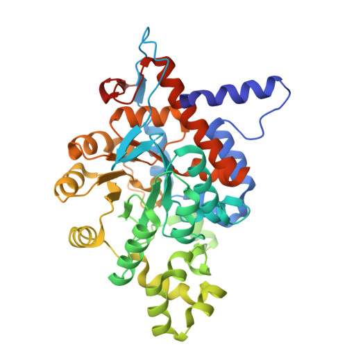
    9EJ8

  • PubMed Abstract: 

    Cross-couplings are essential reactions in modern chemical synthesis, enabling the rapid construction of complex molecules from simple precursors. Transition metal catalysts are prized for these transformations because their reactivity and selectivity can be tuned via judicious selection of the metal and ligand. Although enzymes offer analogous opportunities for tuning via protein engineering, their application to cross-coupling remains limited, as nature relies on alternative paradigms for building molecular complexity. Here, we report the cross-coupling of alkyl halides and benzylic carboxylic acids using an engineered flavin-dependent lactate monooxygenase-a photoenzyme. The enzyme achieves this feat by exploiting the redox versatility of the flavin cofactor. Stoichiometric experiments, ultrafast spectroscopy, and computational studies support a mechanism in which photoexcited flavin quinone initiates the reaction via oxidative decarboxylation to generate a benzylic radical. The resulting flavin semiquinone can reduce the alkyl halide to form a second organic radical within the protein active site, which rapidly engages in C(sp 3 )-C(sp 3 ) bond formation. A variant was engineered to control the stereochemical outcome of this radical-radical coupling event, highlighting the ability of the protein to alter the energetic barrier for a mechanistic step that is traditionally understood to be near barrierless. This work demonstrates that the scope for nonnative reaction mechanisms in biocatalysis far exceeds previously established bounds and has potential to solve a variety of reactivity challenges in cross-coupling chemistry.


  • Organizational Affiliation
    • Department of Chemistry, Princeton University, Princeton, NJ 08544.

Macromolecule Content 

  • Total Structure Weight: 44.79 kDa 
  • Atom Count: 3,670 
  • Modeled Residue Count: 392 
  • Deposited Residue Count: 402 
  • Unique protein chains: 1

Macromolecules

Find similar proteins by:|  3D Structure
Entity ID: 1
MoleculeChains  Sequence LengthOrganismDetailsImage
L-lactate 2-monooxygenase402Mycolicibacterium smegmatisMutation(s): 5 
EC: 1.13.12.4
UniProt
Find proteins for P21795 (Mycolicibacterium smegmatis)
Explore P21795 
Go to UniProtKB:  P21795
Entity Groups
Sequence Clusters30% Identity50% Identity70% Identity90% Identity95% Identity100% Identity
UniProt GroupP21795
Sequence Annotations
Expand
Reference Sequence

Small Molecules

Ligands 4 Unique
IDChains Name / Formula / InChI Key2D Diagram3D Interactions
FMN
(Subject of Investigation/LOI)

Query on FMN


Download:Ideal Coordinates CCD File
B [auth A]FLAVIN MONONUCLEOTIDE
C17 H21 N4 O9 P
FVTCRASFADXXNN-SCRDCRAPSA-N
CIT

Query on CIT


Download:Ideal Coordinates CCD File
D [auth A]CITRIC ACID
C6 H8 O7
KRKNYBCHXYNGOX-UHFFFAOYSA-N
SO4

Query on SO4


Download:Ideal Coordinates CCD File
F [auth A],
G [auth A],
H [auth A]
SULFATE ION
O4 S
QAOWNCQODCNURD-UHFFFAOYSA-L
GOL

Query on GOL


Download:Ideal Coordinates CCD File
C [auth A],
E [auth A]
GLYCEROL
C3 H8 O3
PEDCQBHIVMGVHV-UHFFFAOYSA-N

Experimental Data & Validation

Experimental Data

  • Method: X-RAY DIFFRACTION
  • Resolution: 1.42 Å
  • R-Value Free:  0.178 (Depositor), 0.176 (DCC) 
  • R-Value Work:  0.158 (Depositor), 0.157 (DCC) 
  • R-Value Observed: 0.159 (Depositor) 
Space Group: I 4 2 2
Unit Cell:
Length ( Å )Angle ( ˚ )
a = 110.803α = 90
b = 110.803β = 90
c = 169.659γ = 90
Software Package:
Software NamePurpose
PHENIXrefinement
Aimlessdata scaling
XDSdata reduction
PHASERphasing

Structure Validation

View Full Validation Report


Ligand Structure Quality Assessment 


Entry History 

& Funding Information

Deposition Data


Funding OrganizationLocationGrant Number
National Institutes of Health/National Institute of General Medical Sciences (NIH/NIGMS)United StatesR01 GM127703
H2020 Marie Curie Actions of the European CommissionEuropean Union101063280

Revision History  (Full details and data files)

  • Version 1.0: 2026-02-18
    Type: Initial release