9EI6 | pdb_00009ei6

PasI from Photorhabdus asymbiotica bound to Fe(II) and alpha-ketoglutarate


Experimental Data Snapshot

  • Method: X-RAY DIFFRACTION
  • Resolution: 1.44 Å
  • R-Value Free: 
    0.206 (Depositor), 0.205 (DCC) 
  • R-Value Work: 
    0.173 (Depositor), 0.174 (DCC) 
  • R-Value Observed: 
    0.175 (Depositor) 

Starting Model: in silico
View more details

wwPDB Validation 3D Report Full Report

Validation slider image for 9EI6

Ligand Structure Quality Assessment 


This is version 1.1 of the entry. See complete history

Literature

Unusual O-H Activation-Initiated C-C Bond Cleavage Reaction by a Nonheme Fe Enzyme in Antifungal Nucleoside Biosynthesis.

Du, Y.Dong, J.Draelos, M.M.Collazo-Perez, L.N.Majer, S.H.Boal, A.K.Yokoyama, K.

(2025) J Am Chem Soc 147: 30163-30177

  • DOI: https://doi.org/10.1021/jacs.5c08400
  • Primary Citation Related Structures: 
    9EI6, 9EI7

  • PubMed Abstract: 

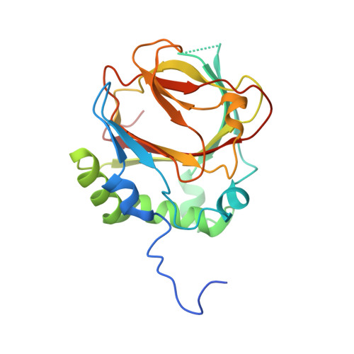
    Fe(II)- and α-ketoglutarate (α-KG)-dependent enzymes catalyze diverse reactions, generally initiated by Fe IV =O mediated cleavage of C-H bonds with bond dissociation energies (BDE) of up to ∼100 kcal/mol. Here, we report the discovery of a novel reaction initiated by a significantly more challenging O-H bond cleavage (>100 kcal/mol). This activity was identified in PolD, an enzyme that regulates the sugar size in antifungal nucleoside biosynthesis by catalyzing the transformation of a bicyclic eight-carbon sugar substrate, 5'-amino-6'-hydroxy-octosyl acid 2'-phosphate (AHOAP), into a monocyclic six-carbon product, aminohexuronic acid 2'-phosphate (AHAP). Our studies demonstrate that PolD catalyzes a two-step reaction, in which AHOAP is first oxidized to 5'-amino-6'-keto-octosyl acid 2'-phosphate (AKOAP) via typical C-H activation, followed by a unique C-C bond cleavage on AKOAP to AHAP initiated by O-H activation. X-ray crystal structures of PolD and its homologue, PasI, the latter solved in complex with AHOAP, succinate, and vanadyl, a structural mimic of the Fe IV -oxo intermediate, reveal a substrate binding mode that is consistent with both C-H and O-H homolysis. A comparison of the three enzymes, PasI, PolD, and MalI, all of which exhibit distinct C-C bond cleavage activities, suggests that precise substrate positioning to bring the target OH group of AKOAP close to the Fe IV -oxo intermediate is critical for hydrogen atom transfer from this functional group. These results indicate a novel reactivity of the Fe IV ═O intermediate in Fe/α-KG enzymes, thereby expanding the reaction scope of this enzyme superfamily. The results also reveal the molecular mechanism of the divergent biosynthesis of antifungal nucleosides.


  • Organizational Affiliation
    • Department of Biochemistry, Duke University School of Medicine, Durham, North Carolina 27710, United States.

Macromolecule Content 

  • Total Structure Weight: 27.6 kDa 
  • Atom Count: 2,074 
  • Modeled Residue Count: 212 
  • Deposited Residue Count: 226 
  • Unique protein chains: 1

Macromolecules

Find similar proteins by:|  3D Structure
Entity ID: 1
MoleculeChains  Sequence LengthOrganismDetailsImage
Prolyl 4-hydroxylase alpha subunit domain-containing protein226Photorhabdus asymbioticaMutation(s): 0 
Gene Names: sanCPAU_00621
UniProt
Find proteins for C7BKM7 (Photorhabdus asymbiotica subsp. asymbiotica (strain ATCC 43949 / 3105-77))
Explore C7BKM7 
Go to UniProtKB:  C7BKM7
Entity Groups
Sequence Clusters30% Identity50% Identity70% Identity90% Identity95% Identity100% Identity
UniProt GroupC7BKM7
Sequence Annotations
Expand
Reference Sequence

Small Molecules

Ligands 5 Unique
IDChains Name / Formula / InChI Key2D Diagram3D Interactions
AKG
(Subject of Investigation/LOI)

Query on AKG


Download:Ideal Coordinates CCD File
B [auth A]2-OXOGLUTARIC ACID
C5 H6 O5
KPGXRSRHYNQIFN-UHFFFAOYSA-N
PEG

Query on PEG


Download:Ideal Coordinates CCD File
G [auth A]DI(HYDROXYETHYL)ETHER
C4 H10 O3
MTHSVFCYNBDYFN-UHFFFAOYSA-N
SO4

Query on SO4


Download:Ideal Coordinates CCD File
C [auth A],
E [auth A],
I [auth A],
J [auth A],
L [auth A]
SULFATE ION
O4 S
QAOWNCQODCNURD-UHFFFAOYSA-L
GOL

Query on GOL


Download:Ideal Coordinates CCD File
D [auth A],
F [auth A],
H [auth A],
K [auth A]
GLYCEROL
C3 H8 O3
PEDCQBHIVMGVHV-UHFFFAOYSA-N
FE
(Subject of Investigation/LOI)

Query on FE


Download:Ideal Coordinates CCD File
M [auth A]FE (III) ION
Fe
VTLYFUHAOXGGBS-UHFFFAOYSA-N

Experimental Data & Validation

Experimental Data

  • Method: X-RAY DIFFRACTION
  • Resolution: 1.44 Å
  • R-Value Free:  0.206 (Depositor), 0.205 (DCC) 
  • R-Value Work:  0.173 (Depositor), 0.174 (DCC) 
  • R-Value Observed: 0.175 (Depositor) 
Space Group: P 21 21 2
Unit Cell:
Length ( Å )Angle ( ˚ )
a = 55.587α = 90
b = 105.336β = 90
c = 39.997γ = 90
Software Package:
Software NamePurpose
PHENIXrefinement
XDSdata scaling
XDSdata reduction
PHENIXphasing

Structure Validation

View Full Validation Report


Ligand Structure Quality Assessment 


Entry History 

& Funding Information

Deposition Data


Funding OrganizationLocationGrant Number
National Institutes of Health/National Institute of General Medical Sciences (NIH/NIGMS)United States5R35GM119707-09

Revision History  (Full details and data files)

  • Version 1.0: 2025-08-20
    Type: Initial release
  • Version 1.1: 2025-08-27
    Changes: Database references