9EF5 | pdb_00009ef5

Cryo-EM structure of Drosophila melanogaster insulin receptor (dmIR) bound with one DILP2, asymmetric conformation


Experimental Data Snapshot

  • Method: ELECTRON MICROSCOPY
  • Resolution: 3.70 Å
  • Aggregation State: PARTICLE 
  • Reconstruction Method: SINGLE PARTICLE 

wwPDB Validation   3D Report Full Report


This is version 1.1 of the entry. See complete history


Literature

Structure and activation of the Drosophila insulin receptor by three Drosophila insulin-like peptides.

Cai, K.Ng, M.Yamamoto, R.R.Hossain, M.A.Hall, C.Wade, J.D.Tatar, M.Choi, E.Bai, X.C.

(2025) Nat Commun 16: 9504-9504

  • DOI: https://doi.org/10.1038/s41467-025-64586-6
  • Primary Citation of Related Structures:  
    9EF1, 9EF4, 9EF5, 9EF9

  • PubMed Abstract: 

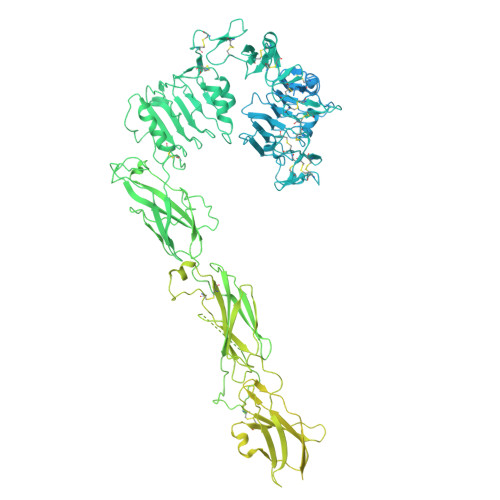
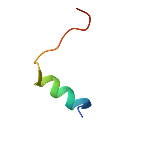
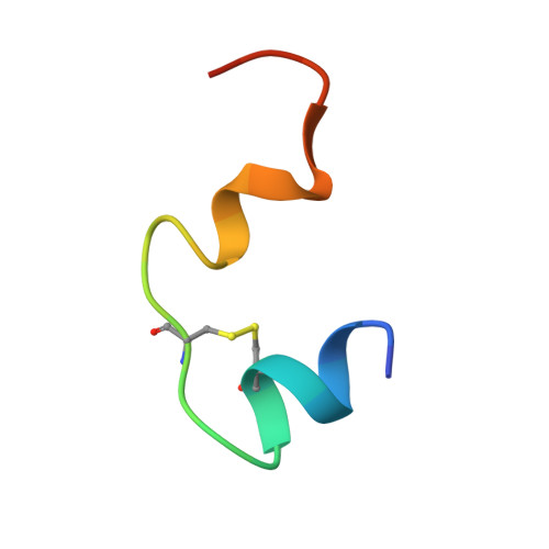
    Insulin/IGF signaling (IIS) is a highly conserved pathway essential for physiological regulation from yeast to mammals. In Drosophila melanogaster, a single insulin-like receptor (dmIR) interacts with various insulin-like peptides (DILPs), leading to diverse signaling and functional outcomes. However, the mechanisms by which different DILPs result in varied receptor activation and biological responses remain unclear. Here, we determine the cryo-electron microscopy (cryo-EM) structures of dmIR in complex with three DILPs: DILP1, DILP2, and DILP5. Our structural analyses reveal that each DILP induces distinct conformations of dmIR: the dmIR/DILP5 complex adopts the Ƭ-shaped asymmetric conformation with three bound DILP5 molecules; the dmIR/DILP2 complex displays the Γ-shaped asymmetric conformation with a single bound DILP2 molecule; and the dmIR/DILP1 complex shows both a Γ-shaped asymmetric conformation and a symmetric conformation that resembles a T-shape with a splayed stem. Functional assays demonstrate that the efficacy of DILP-mediated dmIR activation differs, with DILP5 inducing higher levels of receptor autophosphorylation, followed by DILP2 and DILP1. Together, these findings suggest that the distinct interactions between dmIR and DILPs dictate specific patterns of receptor activation.


  • Organizational Affiliation
    • Department of Biophysics, University of Texas Southwestern Medical Center, Dallas, TX, USA.

Macromolecules
Find similar proteins by:  (by identity cutoff)  |  3D Structure
Entity ID: 1
MoleculeChains Sequence LengthOrganismDetailsImage
Insulin-like receptor
A, B
2,144Drosophila melanogasterMutation(s): 0 
Gene Names: InRDir-aInr-aIRCG18402
EC: 2.7.10.1
UniProt
Find proteins for P09208 (Drosophila melanogaster)
Explore P09208 
Go to UniProtKB:  P09208
Entity Groups  
Sequence Clusters30% Identity50% Identity70% Identity90% Identity95% Identity100% Identity
UniProt GroupP09208
Glycosylation
Glycosylation Sites: 7Go to GlyGen: P09208-1
Sequence Annotations
Expand
  • Reference Sequence

Find similar proteins by:  Sequence   |   3D Structure  

Entity ID: 2
MoleculeChains Sequence LengthOrganismDetailsImage
DILP2 B-chain24Drosophila melanogasterMutation(s): 0 
UniProt
Find proteins for Q9VT51 (Drosophila melanogaster)
Explore Q9VT51 
Go to UniProtKB:  Q9VT51
Entity Groups  
Sequence Clusters30% Identity50% Identity70% Identity90% Identity95% Identity100% Identity
UniProt GroupQ9VT51
Sequence Annotations
Expand
  • Reference Sequence
Find similar proteins by:  (by identity cutoff)  |  3D Structure
Entity ID: 3
MoleculeChains Sequence LengthOrganismDetailsImage
DILP2 A-chain26Drosophila melanogasterMutation(s): 0 
UniProt
Find proteins for Q9VT51 (Drosophila melanogaster)
Explore Q9VT51 
Go to UniProtKB:  Q9VT51
Entity Groups  
Sequence Clusters30% Identity50% Identity70% Identity90% Identity95% Identity100% Identity
UniProt GroupQ9VT51
Sequence Annotations
Expand
  • Reference Sequence
Small Molecules
Ligands 1 Unique
IDChains Name / Formula / InChI Key2D Diagram3D Interactions
NAG (Subject of Investigation/LOI)
Query on NAG

Download Ideal Coordinates CCD File 
E [auth A]
F [auth A]
G [auth A]
H [auth A]
I [auth A]
E [auth A],
F [auth A],
G [auth A],
H [auth A],
I [auth A],
J [auth A],
K [auth A],
L [auth A],
M [auth A],
N [auth B],
O [auth B],
P [auth B],
Q [auth B],
R [auth B],
S [auth B],
T [auth B]
2-acetamido-2-deoxy-beta-D-glucopyranose
C8 H15 N O6
OVRNDRQMDRJTHS-FMDGEEDCSA-N
Experimental Data & Validation

Experimental Data

  • Method: ELECTRON MICROSCOPY
  • Resolution: 3.70 Å
  • Aggregation State: PARTICLE 
  • Reconstruction Method: SINGLE PARTICLE 
EM Software:
TaskSoftware PackageVersion
MODEL REFINEMENTPHENIX
RECONSTRUCTIONRELION

Structure Validation

View Full Validation Report


Entry History & Funding Information

Deposition Data

  • Released Date: 2025-10-01 
  • Deposition Author(s): Bai, X.C.

Funding OrganizationLocationGrant Number
National Institutes of Health/National Institute of General Medical Sciences (NIH/NIGMS)United States--

Revision History  (Full details and data files)

  • Version 1.0: 2025-10-01
    Type: Initial release
  • Version 1.1: 2025-11-12
    Changes: Data collection, Database references