9E83 | pdb_00009e83

TMPRSS2 crystal structure following acylation by UCSF_157


Experimental Data Snapshot

  • Method: X-RAY DIFFRACTION
  • Resolution: 2.07 Å
  • R-Value Free: 
    0.260 (Depositor), 0.265 (DCC) 
  • R-Value Work: 
    0.200 (Depositor), 0.209 (DCC) 
  • R-Value Observed: 
    0.203 (Depositor) 

Starting Model: experimental
View more details

wwPDB Validation   3D Report Full Report


Ligand Structure Quality Assessment 


This is version 1.1 of the entry. See complete history


Literature

Large Library Docking and Biophysical Analysis of Small-Molecule TMPRSS2 Inhibitors.

Fraser, B.J.Young, N.J.Bender, B.J.Gahbauer, S.Ilyassov, O.Wilson, R.P.Li, Y.Seitova, A.Lourenco, A.L.Chung, D.H.Bardine, C.Benard, F.Shoichet, B.K.Craik, C.S.Arrowsmith, C.H.

(2025) J Med Chem 68: 19893-19907

  • DOI: https://doi.org/10.1021/acs.jmedchem.4c03089
  • Primary Citation of Related Structures:  
    8V04, 8V1F, 9E83

  • PubMed Abstract: 

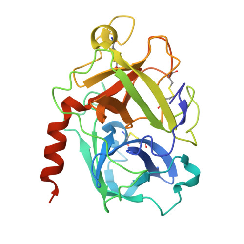
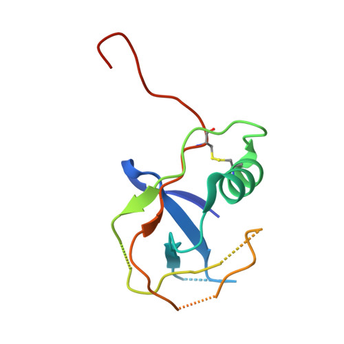
    Transmembrane protease serine-2 (TMPRSS2) is an essential host entry factor in human airways for SARS-CoV-2 and influenza A/B and has presented as a target for antiviral drug development; however, no clinically viable oral small-molecule TMPRSS2 inhibitors have been developed to date. Here, we perform two large-scale docking campaigns to identify covalent and noncovalent TMPRSS2 small-molecule inhibitors using a homology model and crystal structure. We establish a pipeline to rapidly screen TMPRSS2 inhibitors and then interrogate the potency, selectivity, and biophysical properties of covalent and noncovalent inhibition using enzyme kinetics on synthetic peptide and protein substrates and differential scanning fluorimetry. Furthermore, we established a readily crystallizable form of TMPRSS2 protein that produced high-resolution crystal structures with nafamostat , '157 , and 6-amidino-2-naphthol . A novel noncovalent inhibitor scaffold is biochemically validated as a potential avenue for developing TMPRSS2-selective inhibitors.


  • Organizational Affiliation
    • Structural Genomics Consortium Toronto, Toronto, Ontario M5G 1L7, Canada.

Macromolecules
Find similar proteins by:  (by identity cutoff)  |  3D Structure
Entity ID: 1
MoleculeChains Sequence LengthOrganismDetailsImage
Transmembrane protease serine 2A [auth B]249Homo sapiensMutation(s): 0 
Gene Names: TMPRSS2
EC: 3.4.21.122
UniProt & NIH Common Fund Data Resources
Find proteins for O15393 (Homo sapiens)
Explore O15393 
Go to UniProtKB:  O15393
PHAROS:  O15393
GTEx:  ENSG00000184012 
Entity Groups  
Sequence Clusters30% Identity50% Identity70% Identity90% Identity95% Identity100% Identity
UniProt GroupO15393
Sequence Annotations
Expand
  • Reference Sequence
Find similar proteins by:  (by identity cutoff)  |  3D Structure
Entity ID: 2
MoleculeChains Sequence LengthOrganismDetailsImage
Transmembrane protease serine 2 non-catalytic chainB [auth A]110Homo sapiensMutation(s): 6 
Gene Names: TMPRSS2PRSS10
EC: 3.4.21.122
UniProt & NIH Common Fund Data Resources
Find proteins for O15393 (Homo sapiens)
Explore O15393 
Go to UniProtKB:  O15393
PHAROS:  O15393
GTEx:  ENSG00000184012 
Entity Groups  
Sequence Clusters30% Identity50% Identity70% Identity90% Identity95% Identity100% Identity
UniProt GroupO15393
Sequence Annotations
Expand
  • Reference Sequence
Small Molecules
Ligands 4 Unique
IDChains Name / Formula / InChI Key2D Diagram3D Interactions
CIT
Query on CIT

Download Ideal Coordinates CCD File 
C [auth B]CITRIC ACID
C6 H8 O7
KRKNYBCHXYNGOX-UHFFFAOYSA-N
VU4 (Subject of Investigation/LOI)
Query on VU4

Download Ideal Coordinates CCD File 
E [auth B]4-(2-aminoethyl)benzoic acid
C9 H11 N O2
MRBFWTDIRYEDBQ-UHFFFAOYSA-N
EDO
Query on EDO

Download Ideal Coordinates CCD File 
D [auth B],
F [auth B],
G [auth B]
1,2-ETHANEDIOL
C2 H6 O2
LYCAIKOWRPUZTN-UHFFFAOYSA-N
UNX (Subject of Investigation/LOI)
Query on UNX

Download Ideal Coordinates CCD File 
H [auth A]UNKNOWN ATOM OR ION
X
Experimental Data & Validation

Experimental Data

  • Method: X-RAY DIFFRACTION
  • Resolution: 2.07 Å
  • R-Value Free:  0.260 (Depositor), 0.265 (DCC) 
  • R-Value Work:  0.200 (Depositor), 0.209 (DCC) 
  • R-Value Observed: 0.203 (Depositor) 
Space Group: P 1 21 1
Unit Cell:
Length ( Å )Angle ( ˚ )
a = 60.853α = 90
b = 51.392β = 91.01
c = 64.625γ = 90
Software Package:
Software NamePurpose
REFMACrefinement
HKL-3000data scaling
HKL-3000data reduction
PHASERphasing

Structure Validation

View Full Validation Report


Ligand Structure Quality Assessment 


Entry History & Funding Information

Deposition Data


Funding OrganizationLocationGrant Number
Canadian Institutes of Health Research (CIHR)CanadaFDN154328
National Institutes of Health/National Institute of General Medical Sciences (NIH/NIGMS)United StatesP30 GM124165

Revision History  (Full details and data files)

  • Version 1.0: 2024-12-25
    Type: Initial release
  • Version 1.1: 2026-01-14
    Changes: Database references, Derived calculations, Structure summary