9E6H | pdb_00009e6h

Cryo-EM structure of Maackia amurensis seed Leukoagglutinin (lectin), MASL


Experimental Data Snapshot

  • Method: ELECTRON MICROSCOPY
  • Resolution: 2.84 Å
  • Aggregation State: PARTICLE 
  • Reconstruction Method: SINGLE PARTICLE 

Starting Model: experimental
View more details

wwPDB Validation   3D Report Full Report


This is version 1.2 of the entry. See complete history


Literature

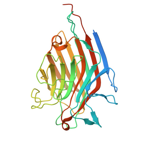
Maackia amurensis seed lectin structure and sequence comparison with other M. amurensis lectins.

Nayak, A.R.Holdcraft, C.J.Yin, A.C.Nicoletto, R.E.Zhao, C.Zheng, H.Temiakov, D.Goldberg, G.S.

(2025) J Biological Chem 301: 108466-108466

  • DOI: https://doi.org/10.1016/j.jbc.2025.108466
  • Primary Citation of Related Structures:  
    9E6H

  • PubMed Abstract: 

    Maackia amurensis lectins, including MASL, MAA, and MAL2, are widely utilized in biochemical and medicinal research. However, the structural and functional differences between these lectins have not been defined. Here, we present a high-resolution cryo-EM structure of MASL revealing that its tetrameric assembly is directed by two intersubunit disulfide bridges. These bridges, formed by C272 residues, are central to the dimer-of-dimers assembly of a MASL tetramer. This cryo-EM structure also identifies residues involved in stabilizing the dimer interface, multiple glycosylation sites, and calcium and manganese atoms in the sugar-binding pockets of MASL. Notably, our analysis reveals that Y250 in the carbohydrate-binding site of MASL adopts a flipped conformation, likely acting as a gatekeeper that obstructs access to non-cognate substrates, a feature that may contribute to MASL's substrate specificity. Sequence analysis suggests that MAA is a truncated version of MASL, while MAL2 represents a homologous isoform. Unlike MASL, neither MAL2 nor MAA contains a cysteine residue required for disulfide bridge formation. Accordingly, analysis of these proteins using reducing and nonreducing SDS-PAGE confirms that the C272 residue in MASL drives intermolecular disulfide bridge formation. These findings provide critical insights into the unique structural features of MASL that distinguish it from other Maackia amurensis lectins, offering a foundation for further exploration of its biological and therapeutic potential.


  • Organizational Affiliation
    • Biochemistry & Molecular Biology Department, Thomas Jefferson University, 1020 Locust Street, Philadelphia, PA 19107, USA.

Macromolecules
Find similar proteins by:  (by identity cutoff)  |  3D Structure
Entity ID: 1
MoleculeChains Sequence LengthOrganismDetailsImage
Seed leukoagglutinin
A, B, C, D
287Maackia amurensisMutation(s): 0 
UniProt
Find proteins for P0DKL3 (Maackia amurensis)
Explore P0DKL3 
Go to UniProtKB:  P0DKL3
Entity Groups  
Sequence Clusters30% Identity50% Identity70% Identity90% Identity95% Identity100% Identity
UniProt GroupP0DKL3
Glycosylation
Glycosylation Sites: 4
Sequence Annotations
Expand
  • Reference Sequence
Oligosaccharides

Help

Entity ID: 2
MoleculeChains Length2D Diagram Glycosylation3D Interactions
alpha-D-mannopyranose-(1-3)-beta-D-mannopyranose-(1-4)-2-acetamido-2-deoxy-beta-D-glucopyranose-(1-4)-2-acetamido-2-deoxy-beta-D-glucopyranose
E, G, I, K
4N-Glycosylation
Glycosylation Resources
GlyTouCan:  G81315DD
GlyCosmos:  G81315DD
GlyGen:  G81315DD
Entity ID: 3
MoleculeChains Length2D Diagram Glycosylation3D Interactions
alpha-D-mannopyranose-(1-3)-[alpha-D-mannopyranose-(1-6)]beta-D-mannopyranose-(1-4)-2-acetamido-2-deoxy-beta-D-glucopyranose-(1-4)-2-acetamido-2-deoxy-beta-D-glucopyranose
F, H, J, L
5N-Glycosylation
Glycosylation Resources
GlyTouCan:  G22768VO
GlyCosmos:  G22768VO
GlyGen:  G22768VO
Small Molecules
Ligands 3 Unique
IDChains Name / Formula / InChI Key2D Diagram3D Interactions
NAG (Subject of Investigation/LOI)
Query on NAG

Download Ideal Coordinates CCD File 
M [auth A]
N [auth A]
Q [auth B]
R [auth B]
U [auth C]
M [auth A],
N [auth A],
Q [auth B],
R [auth B],
U [auth C],
V [auth C],
Y [auth D],
Z [auth D]
2-acetamido-2-deoxy-beta-D-glucopyranose
C8 H15 N O6
OVRNDRQMDRJTHS-FMDGEEDCSA-N
MN
Query on MN

Download Ideal Coordinates CCD File 
BA [auth D],
P [auth A],
T [auth B],
X [auth C]
MANGANESE (II) ION
Mn
WAEMQWOKJMHJLA-UHFFFAOYSA-N
CA
Query on CA

Download Ideal Coordinates CCD File 
AA [auth D],
O [auth A],
S [auth B],
W [auth C]
CALCIUM ION
Ca
BHPQYMZQTOCNFJ-UHFFFAOYSA-N
Experimental Data & Validation

Experimental Data

  • Method: ELECTRON MICROSCOPY
  • Resolution: 2.84 Å
  • Aggregation State: PARTICLE 
  • Reconstruction Method: SINGLE PARTICLE 
EM Software:
TaskSoftware PackageVersion
MODEL REFINEMENTPHENIX1.21.2.5419
RECONSTRUCTIONcryoSPARC4.4

Structure Validation

View Full Validation Report


Entry History & Funding Information

Deposition Data


Funding OrganizationLocationGrant Number
National Institutes of Health/National Institute of General Medical Sciences (NIH/NIGMS)United StatesNIH GM131832

Revision History  (Full details and data files)

  • Version 1.0: 2025-04-09
    Type: Initial release
  • Version 1.1: 2025-04-16
    Changes: Data collection, Database references
  • Version 1.2: 2025-05-07
    Changes: Data collection, Database references