9E3G | pdb_00009e3g

Torpedo muscle-type nicotinic acetylcholine receptor - Monoliganded State


Experimental Data Snapshot

  • Method: ELECTRON MICROSCOPY
  • Resolution: 3.13 Å
  • Aggregation State: PARTICLE 
  • Reconstruction Method: SINGLE PARTICLE 

wwPDB Validation 3D Report Full Report

Validation slider image for 9E3G

This is version 2.0 of the entry. See complete history

Literature

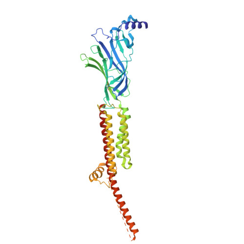
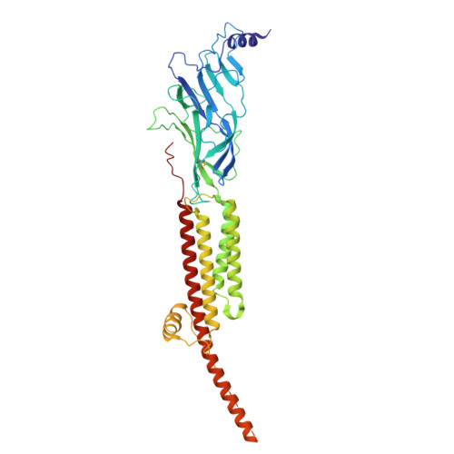
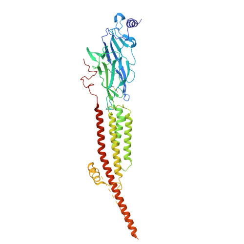
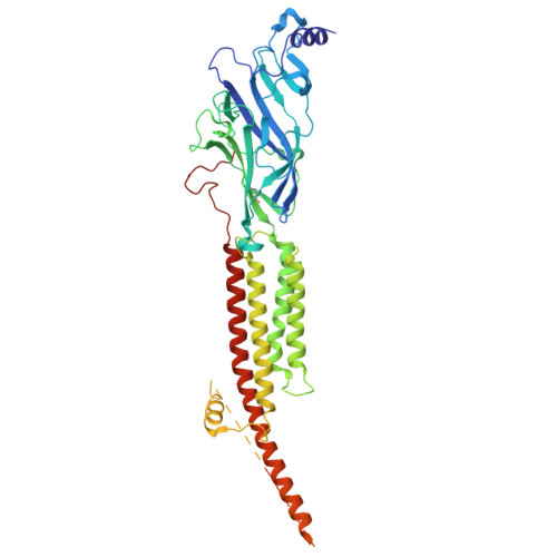
Asynchronous subunit transitions prime acetylcholine receptor activation.

Thompson, M.J.Tessier, C.J.G.Ananchenko, A.Henault, C.Emlaw, J.R.Dehez, F.Zarkadas, E.daCosta, C.J.B.Nury, H.Baenziger, J.E.

(2026) Science 391: eadw1264-eadw1264

  • DOI: https://doi.org/10.1126/science.adw1264
  • Primary Citation Related Structures: 
    9E3E, 9E3F, 9E3G

  • PubMed Abstract: 

    Communication at synapses is facilitated by postsynaptic receptors, which convert a chemical signal into an electrical response. For ligand-gated ion channels, agonist binding triggers rapid transitions through intermediate states leading to a transient open-pore conformation, with these transitions shaping the post-synaptic response. Here, we determine structures of the muscle-type nicotinic acetylcholine receptor in unliganded, mono-liganded, and di-liganded states. Agonist binding to a single site stabilizes a closed structure where an entire principal agonist-binding subunit transitions to an active-like conformation, while the other unoccupied principal subunit remains inactive, albeit poised for activation. Uniting this intermediate structure with single-channel recordings informs a sequential activation mechanism where asynchronous subunit transitions prime the receptor for activation, a finding with implications for an entire superfamily of pentameric ligand-gated ion channels.


  • Organizational Affiliation
    • Department of Biochemistry, Microbiology, and Immunology, University of Ottawa, Ottawa, ON, Canada.

Macromolecule Content 

  • Total Structure Weight: 278.55 kDa 
  • Atom Count: 16,980 
  • Modeled Residue Count: 2,013 
  • Deposited Residue Count: 2,332 
  • Unique protein chains: 4

Macromolecules

Find similar proteins by:|  3D Structure
Entity ID: 1
MoleculeChains  Sequence LengthOrganismDetailsImage
Acetylcholine receptor subunit alpha
A, D
437Tetronarce californicaMutation(s): 0 
UniProt
Find proteins for P02710 (Tetronarce californica)
Explore P02710 
Go to UniProtKB:  P02710
Entity Groups
Sequence Clusters30% Identity50% Identity70% Identity90% Identity95% Identity100% Identity
UniProt GroupP02710
Glycosylation
Glycosylation Sites: 1
Sequence Annotations
Expand
Reference Sequence
Find similar proteins by:|  3D Structure
Entity ID: 2
MoleculeChains  Sequence LengthOrganismDetailsImage
Acetylcholine receptor subunit beta469Tetronarce californicaMutation(s): 0 
UniProt
Find proteins for P02712 (Tetronarce californica)
Explore P02712 
Go to UniProtKB:  P02712
Entity Groups
Sequence Clusters30% Identity50% Identity70% Identity90% Identity95% Identity100% Identity
UniProt GroupP02712
Glycosylation
Glycosylation Sites: 1
Sequence Annotations
Expand
Reference Sequence
Find similar proteins by:|  3D Structure
Entity ID: 3
MoleculeChains  Sequence LengthOrganismDetailsImage
Acetylcholine receptor subunit delta501Tetronarce californicaMutation(s): 0 
UniProt
Find proteins for P02718 (Tetronarce californica)
Explore P02718 
Go to UniProtKB:  P02718
Entity Groups
Sequence Clusters30% Identity50% Identity70% Identity90% Identity95% Identity100% Identity
UniProt GroupP02718
Glycosylation
Glycosylation Sites: 3
Sequence Annotations
Expand
Reference Sequence
Find similar proteins by:|  3D Structure
Entity ID: 4
MoleculeChains  Sequence LengthOrganismDetailsImage
Acetylcholine receptor subunit gamma488Tetronarce californicaMutation(s): 0 
UniProt
Find proteins for P02714 (Tetronarce californica)
Explore P02714 
Go to UniProtKB:  P02714
Entity Groups
Sequence Clusters30% Identity50% Identity70% Identity90% Identity95% Identity100% Identity
UniProt GroupP02714
Glycosylation
Glycosylation Sites: 1
Sequence Annotations
Expand
Reference Sequence

Oligosaccharides

Help  
Entity ID: 5
MoleculeChains Length2D Diagram GlycosylationD Interactions
alpha-D-mannopyranose-(1-3)-[alpha-D-mannopyranose-(1-6)]alpha-D-mannopyranose-(1-6)-[alpha-D-mannopyranose-(1-3)]beta-D-mannopyranose-(1-4)-2-acetamido-2-deoxy-beta-D-glucopyranose-(1-4)-2-acetamido-2-deoxy-beta-D-glucopyranose
F
7N-Glycosylation
Glycosylation Resources
GlyTouCan: G55220VL
GlyCosmos: G55220VL
GlyGen: G55220VL
Entity ID: 6
MoleculeChains Length2D Diagram GlycosylationD Interactions
alpha-D-mannopyranose-(1-3)-[alpha-D-mannopyranose-(1-6)]beta-D-mannopyranose-(1-4)-2-acetamido-2-deoxy-beta-D-glucopyranose-(1-4)-2-acetamido-2-deoxy-beta-D-glucopyranose
G, H
5N-Glycosylation
Glycosylation Resources
GlyTouCan: G22768VO
GlyCosmos: G22768VO
GlyGen: G22768VO
Entity ID: 7
MoleculeChains Length2D Diagram GlycosylationD Interactions
alpha-D-mannopyranose-(1-2)-alpha-D-mannopyranose-(1-3)-[alpha-D-mannopyranose-(1-6)]beta-D-mannopyranose-(1-4)-2-acetamido-2-deoxy-beta-D-glucopyranose-(1-4)-2-acetamido-2-deoxy-beta-D-glucopyranose
I
6N-Glycosylation
Glycosylation Resources
GlyTouCan: G56014GC
GlyCosmos: G56014GC
GlyGen: G56014GC
Entity ID: 8
MoleculeChains Length2D Diagram GlycosylationD Interactions
2-acetamido-2-deoxy-beta-D-glucopyranose-(1-4)-2-acetamido-2-deoxy-beta-D-glucopyranose
J
2N-Glycosylation
Glycosylation Resources
GlyTouCan: G42666HT
GlyCosmos: G42666HT
GlyGen: G42666HT
Entity ID: 9
MoleculeChains Length2D Diagram GlycosylationD Interactions
alpha-D-mannopyranose-(1-6)-alpha-D-mannopyranose-(1-6)-[alpha-D-mannopyranose-(1-3)]beta-D-mannopyranose-(1-4)-2-acetamido-2-deoxy-beta-D-glucopyranose-(1-4)-2-acetamido-2-deoxy-beta-D-glucopyranose
K, L
6N-Glycosylation
Glycosylation Resources
GlyTouCan: G34442SS
GlyCosmos: G34442SS
GlyGen: G34442SS

Experimental Data & Validation

Experimental Data

  • Method: ELECTRON MICROSCOPY
  • Resolution: 3.13 Å
  • Aggregation State: PARTICLE 
  • Reconstruction Method: SINGLE PARTICLE 
EM Software:
TaskSoftware PackageVersion
MODEL REFINEMENTPHENIX1.21rc1_4958
RECONSTRUCTIONcryoSPARC3.9

Structure Validation

View Full Validation Report


Entry History 

& Funding Information

Deposition Data


Funding OrganizationLocationGrant Number
Canadian Institutes of Health Research (CIHR)Canada175223
Natural Sciences and Engineering Research Council (NSERC, Canada)CanadaRGPIN-2022-04723
European Research Council (ERC)European Union637733
Natural Sciences and Engineering Research Council (NSERC, Canada)CanadaRGPIN-2016-04801
Canada Foundation for InnovationCanada34475
Canadian Institutes of Health Research (CIHR)Canada377068

Revision History  (Full details and data files)

  • Version 1.0: 2025-10-08
    Type: Initial release
  • Version 1.1: 2025-10-15
    Changes: Data collection, Database references
  • Version 2.0: 2026-01-14
    Type: Coordinate replacement
    Reason: Model orientation/position
    Changes: Advisory, Atomic model, Author supporting evidence, Data collection, Database references, Derived calculations, Refinement description, Structure summary