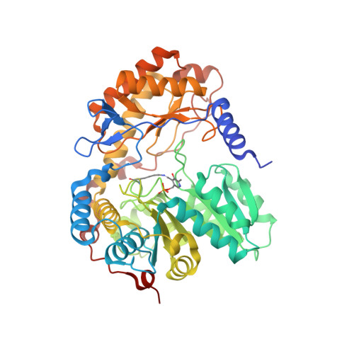
9DZO | pdb_00009dzo

Structure of ALAS bound to succinyl-CoA from S. cerevisiae


Experimental Data Snapshot

  • Method: X-RAY DIFFRACTION
  • Resolution: 2.69 Å
  • R-Value Free: 
    0.227 (Depositor), 0.231 (DCC) 
  • R-Value Work: 
    0.180 (Depositor), 0.186 (DCC) 

Starting Model: experimental
View more details

wwPDB Validation 3D Report Full Report

Validation slider image for 9DZO

Ligand Structure Quality Assessment 


This is version 1.1 of the entry. See complete history. 

Literature

Substrate-bound structures of yeast ALA synthase reveal protein dynamics initiating heme biosynthesis.

Tran, J.U., Martinez, T.A., Jurich, C., Josling, M.R., Zhang, J.M., Ayres-Galhardo, P.H., Meiler, J., Yang, Z.J., MacGurn, J.A., Brown, B.L.

(2026) Sci Adv 12: eaef7768-eaef7768

  • DOI: https://doi.org/10.1126/sciadv.aef7768
  • Primary Citation Related Structures: 
    9DZ7, 9DZO

  • PubMed Abstract: 

    Heme is an essential prosthetic group involved in multiple biological processes in nearly all organisms. Heme biosynthesis is initiated by the pyridoxal 5'-phosphate-dependent enzyme aminolevulinic acid synthase (ALAS). Mutations in ALAS cause multiple hematological diseases. Despite sharing approximately 70% sequence similarity, ALAS homologs exhibit distinct expression and regulatory mechanisms. However, the molecular basis for their functional divergence remains poorly understood. Here, we used x-ray crystallography and molecular dynamics simulations to elucidate the structure and dynamics of Saccharomyces cerevisiae ALAS (scALAS) upon substrate binding. We identified critical conformational changes that occur during catalysis, regulating enzyme activity and protein-protein interactions specific to eukaryotic heme synthesis. Using evolution-guided engineering, we generated scALAS variants with altered catalytic activity and cellular metabolic profiles. Our findings reveal molecular and structural mechanisms underlying heme synthesis regulation and establish a framework for developing targeted therapeutics to treat heme-deficiency disorders and inform a rational design of next-generation therapeutic tools.


  • Organizational Affiliation: 
    • Department of Biochemistry, Vanderbilt University School of Medicine, Nashville, TN, USA.

Macromolecule Content 

  • Total Structure Weight: 329.65 kDa 
  • Atom Count: 23,215 
  • Modeled Residue Count: 2,865 
  • Deposited Residue Count: 2,946 
  • Unique protein chains: 2

Macromolecules

Find similar proteins by:|  3D Structure
Entity ID: 1
MoleculeChains  Sequence LengthOrganismDetailsImage
5-aminolevulinate synthase, mitochondrialA [auth C],
B [auth D],
C [auth A],
D [auth E],
E [auth B]
491Saccharomyces cerevisiaeMutation(s): 0 
Gene Names: HEM1, CYD1, YDR232W, YD9934.16
EC: 2.3.1.37
UniProt
Find proteins for P09950 (Saccharomyces cerevisiae (strain ATCC 204508 / S288c))
Explore P09950 
Go to UniProtKB:  P09950
Entity Groups
Sequence Clusters30% Identity50% Identity70% Identity90% Identity95% Identity100% Identity
UniProt GroupP09950
Sequence Annotations
Expand
Reference Sequence
Find similar proteins by:|  3D Structure
Entity ID: 2
MoleculeChains  Sequence LengthOrganismDetailsImage
5-aminolevulinate synthase, mitochondrial491Saccharomyces cerevisiaeMutation(s): 0 
Gene Names: HEM1, CYD1, YDR232W, YD9934.16
EC: 2.3.1.37
UniProt
Find proteins for P09950 (Saccharomyces cerevisiae (strain ATCC 204508 / S288c))
Explore P09950 
Go to UniProtKB:  P09950
Entity Groups
Sequence Clusters30% Identity50% Identity70% Identity90% Identity95% Identity100% Identity
UniProt GroupP09950
Sequence Annotations
Expand
Reference Sequence

Small Molecules

Ligands 3 Unique
IDChains Name / Formula / InChI Key2D Diagram3D Interactions
SCA
(Subject of Investigation/LOI)

Query on SCA


Download:Ideal Coordinates CCD File
H [auth C],
L [auth A],
N [auth E],
P [auth B],
Q [auth F]
SUCCINYL-COENZYME A
C25 H40 N7 O19 P3 S
VNOYUJKHFWYWIR-ITIYDSSPSA-N
PLP
(Subject of Investigation/LOI)

Query on PLP


Download:Ideal Coordinates CCD File
G [auth C],
I [auth D],
K [auth A],
M [auth E],
O [auth B]
PYRIDOXAL-5'-PHOSPHATE
C8 H10 N O6 P
NGVDGCNFYWLIFO-UHFFFAOYSA-N
MG

Query on MG


Download:Ideal Coordinates CCD File
J [auth D]MAGNESIUM ION
Mg
JLVVSXFLKOJNIY-UHFFFAOYSA-N
Modified Residues  1 Unique
IDChains Modified ResidueModification TypeFormula2D Diagram
LLP
Query on LLP
LYSPyridoxal phosphateC14 H22 N3 O7 P

Experimental Data & Validation

Experimental Data

  • Method: X-RAY DIFFRACTION
  • Resolution: 2.69 Å
  • R-Value Free:  0.227 (Depositor), 0.231 (DCC) 
  • R-Value Work:  0.180 (Depositor), 0.186 (DCC) 
Space Group: P 1
Unit Cell:
Length ( Å )Angle ( ˚ )
a = 60.595α = 116.403
b = 114.241β = 97.507
c = 119.307γ = 91.839
Software Package:
Software NamePurpose
REFMACrefinement
XDSdata reduction
XDSdata scaling
PHASERphasing

Structure Validation

View Full Validation Report


Ligand Structure Quality Assessment 


Entry History 

& Funding Information

Deposition Data


Funding OrganizationLocationGrant Number
National Institutes of Health/National Institute of General Medical Sciences (NIH/NIGMS)United StatesDP2 GM146255
American Heart AssociationUnited States24PRE1190012

Revision History  (Full details and data files)

  • Version 1.0: 2025-10-22
    Type: Initial release
  • Version 1.1: 2026-10-28
    Changes: Database references