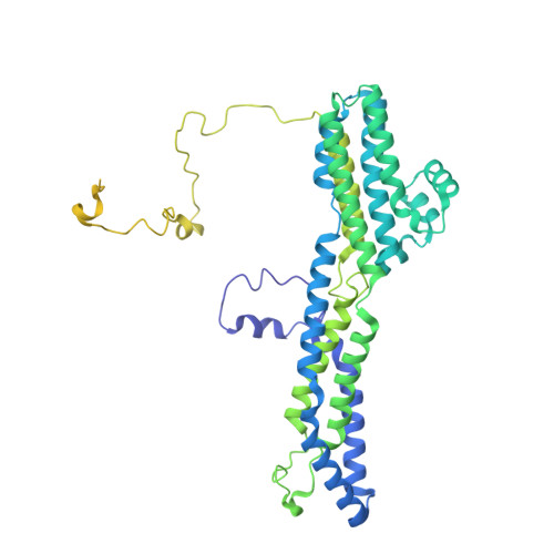
Neurotransmitter-bound bestrophin channel structures reveal small molecule drug targeting sites for disease treatment.
Owji, A.P., Dong, J., Kittredge, A., Wang, J., Zhang, Y., Yang, T.(2024) Nat Commun 15: 10766-10766
- PubMed: 39737942
- DOI: https://doi.org/10.1038/s41467-024-54938-z
- Primary Citation of Related Structures:
9DYH, 9DYI, 9DYJ, 9DYK, 9DYL, 9DYM, 9DYN, 9DYO - PubMed Abstract:
Best1 and Best2 are two members of the bestrophin family of anion channels critically involved in the prevention of retinal degeneration and maintenance of intraocular pressure, respectively. Here, we solved glutamate- and γ-aminobutyric acid (GABA)-bound Best2 structures, which delineate an intracellular glutamate binding site and an extracellular GABA binding site on Best2, respectively, identified extracellular GABA as a permeable activator of Best2, and elucidated the co-regulation of Best2 by glutamate, GABA and glutamine synthetase in vivo. We further identified multiple small molecules as activators of the bestrophin channels. Extensive analyses were carried out for a potent activator, 4-aminobenzoic acid (PABA): PABA-bound Best1 and Best2 structures are solved and illustrate the same binding site as in GABA-bound Best2; PABA treatment rescues the functional deficiency of patient-derived Best1 mutations. Together, our results demonstrate the mechanism and potential of multiple small molecule candidates as clinically applicable drugs for bestrophin-associated diseases/conditions.
- Department of Ophthalmology, Columbia University, New York, NY, USA.
Organizational Affiliation:


