9DVI | pdb_00009dvi

Hetero-hexameric Hsp70-Hsp40-DafA complex


Experimental Data Snapshot

  • Method: ELECTRON MICROSCOPY
  • Resolution: 3.37 Å
  • Aggregation State: PARTICLE 
  • Reconstruction Method: SINGLE PARTICLE 

wwPDB Validation 3D Report Full Report

Validation slider image for 9DVI

This is version 1.1 of the entry. See complete history

Literature

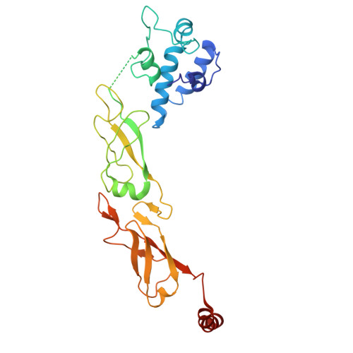
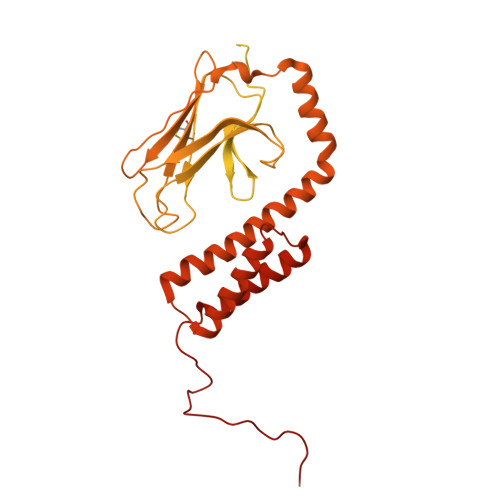
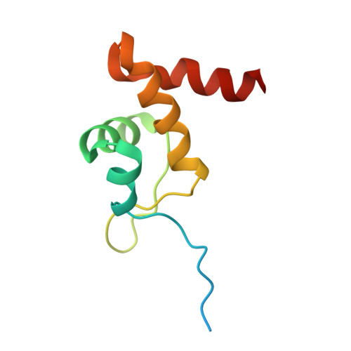
Mechanisms of assembly and function of the Hsp70-Hsp40 chaperone machinery.

Jiang, Y.Ibrahim, Z.Xia, Y.Clay, M.Myasnikov, A.Immadisetty, K.Xia, Z.Tang, L.Rossi, P.Ganguly, P.Liu, J.Miller, D.Che, M.Palacios, S.M.Kramer, G.Bukau, B.Kalodimos, C.G.

(2025) Mol Cell 85: 4032-4046.e7

  • DOI: https://doi.org/10.1016/j.molcel.2025.09.023
  • Primary Citation Related Structures: 
    8W1M, 9DVI, 9DYU, 9DYV

  • PubMed Abstract: 

    Hsp70 and Hsp40 molecular chaperones form a central machinery that remodels client proteins involved in numerous biological processes. Here, we integrated cryo-electron microscopy and nuclear magnetic resonance spectroscopy to determine the architecture of the full-length Hsp70-Hsp40 machinery. The structure of the complex in a physiologically inhibited state reveals distinct regulatory mechanisms. In the active state, the Hsp40 glycine-phenylalanine (G/F)-rich region acts as a pseudo-substrate for Hsp70, directly modulating refolding. This region also maintains Hsp40 in an autoinhibited state; upon binding to Hsp70, the inhibition is disrupted, exposing a cryptic client-binding site that enables client engagement and refolding. Transitions between these states are central to controlling refolding efficiency. Disrupting either the autoinhibited state or the G/F-Hsp70 interaction impairs function and elicits a compensatory heat shock response in cells. Our findings uncover the regulatory dynamics of a fundamental chaperone system, with broad implications for understanding protein homeostasis and the cellular response to stress.


  • Organizational Affiliation
    • State Key Laboratory of Pharmaceutical Biotechnology, Department of Oncology, Nanjing Drum Tower Hospital, School of Life Sciences, Chemistry and Biomedicine Innovation Center (ChemBIC), Nanjing University, Nanjing, China; Department of Structural Biology, St. Jude Children's Research Hospital, Memphis, TN, USA.

Macromolecule Content 

  • Total Structure Weight: 216.8 kDa 
  • Atom Count: 8,978 
  • Modeled Residue Count: 1,153 
  • Deposited Residue Count: 1,974 
  • Unique protein chains: 3

Macromolecules

Find similar proteins by:|  3D Structure
Entity ID: 1
MoleculeChains  Sequence LengthOrganismDetailsImage
Chaperone protein DnaJ 2
A, C
280Thermus thermophilusMutation(s): 0 
Gene Names: dnaJ2TthHB5018_15880
UniProt
Find proteins for Q72IK7 (Thermus thermophilus (strain ATCC BAA-163 / DSM 7039 / HB27))
Explore Q72IK7 
Go to UniProtKB:  Q72IK7
Entity Groups
Sequence Clusters30% Identity50% Identity70% Identity90% Identity95% Identity100% Identity
UniProt GroupQ72IK7
Sequence Annotations
Expand
Reference Sequence
Find similar proteins by:|  3D Structure
Entity ID: 2
MoleculeChains  Sequence LengthOrganismDetailsImage
Chaperone protein DnaK
B, D
615Thermus thermophilusMutation(s): 0 
Gene Names: dnaKTthHB5018_15900
UniProt
Find proteins for Q72IK5 (Thermus thermophilus (strain ATCC BAA-163 / DSM 7039 / HB27))
Explore Q72IK5 
Go to UniProtKB:  Q72IK5
Entity Groups
Sequence Clusters30% Identity50% Identity70% Identity90% Identity95% Identity100% Identity
UniProt GroupQ72IK5
Sequence Annotations
Expand
Reference Sequence
Find similar proteins by:|  3D Structure
Entity ID: 3
MoleculeChains  Sequence LengthOrganismDetailsImage
Protein DafAE [auth S],
F [auth T]
92Thermus thermophilusMutation(s): 0 
Gene Names: dafATthAA11_16070TthHB5018_15870
UniProt
Find proteins for Q72IK8 (Thermus thermophilus (strain ATCC BAA-163 / DSM 7039 / HB27))
Explore Q72IK8 
Go to UniProtKB:  Q72IK8
Entity Groups
Sequence Clusters30% Identity50% Identity70% Identity90% Identity95% Identity100% Identity
UniProt GroupQ72IK8
Sequence Annotations
Expand
Reference Sequence

Experimental Data & Validation

Experimental Data

  • Method: ELECTRON MICROSCOPY
  • Resolution: 3.37 Å
  • Aggregation State: PARTICLE 
  • Reconstruction Method: SINGLE PARTICLE 
EM Software:
TaskSoftware PackageVersion
MODEL REFINEMENTPHENIX1.21_5207:

Structure Validation

View Full Validation Report


Entry History 

& Funding Information

Deposition Data


Funding OrganizationLocationGrant Number
Other governmentUnited StatesNIH grant R35 GM12246
Other privateUnited StatesALSAC

Revision History  (Full details and data files)

  • Version 1.0: 2025-10-22
    Type: Initial release
  • Version 1.1: 2026-05-06
    Changes: Database references