9DVA

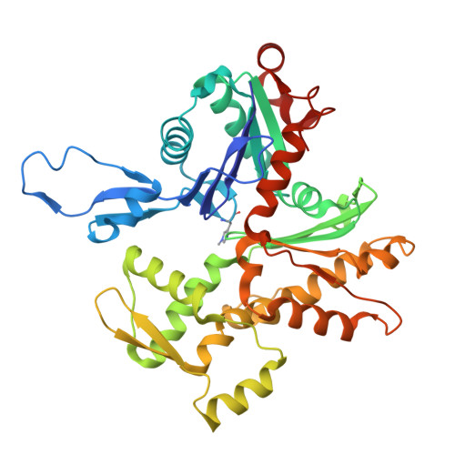
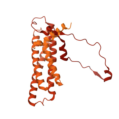
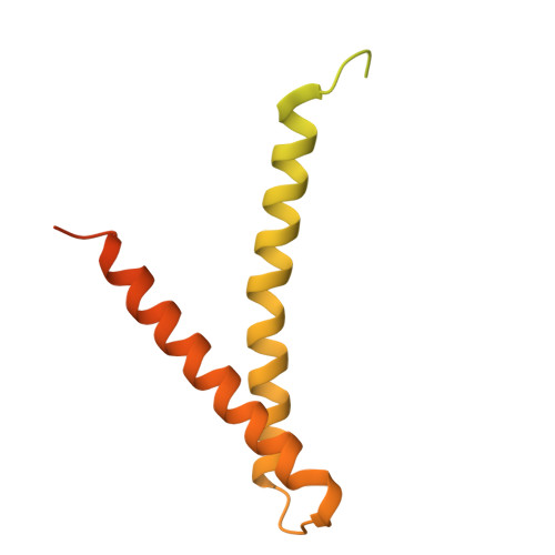
F-actin binding interface of alpha-E-catenin ABD (cadherin-catenin complex) and afadin


Experimental Data Snapshot

  • Method: ELECTRON MICROSCOPY
  • Resolution: 3.10 Å
  • Aggregation State: FILAMENT 
  • Reconstruction Method: SINGLE PARTICLE 

wwPDB Validation   3D Report Full Report


This is version 1.0 of the entry. See complete history


Literature

Afadin mediates cadherin-catenin complex clustering on F-actin linked to cooperative binding and filament curvature.

Gong, R.Reynolds, M.J.Sun, X.Alushin, G.M.

(2024) bioRxiv 

  • DOI: https://doi.org/10.1101/2024.10.08.617332
  • Primary Citation of Related Structures:  
    9DVA

  • PubMed Abstract: 

    The E-cadherin-β-catenin-αE-catenin (cadherin-catenin) complex couples the cytoskeletons of neighboring cells at adherens junctions (AJs) to mediate force transmission across epithelia. Mechanical force and auxiliary binding partners converge to stabilize the cadherin-catenin complex's inherently weak binding to actin filaments (F-actin) through unclear mechanisms. Here we show that afadin's coiled-coil (CC) domain and vinculin synergistically enhance the cadherin-catenin complex's F-actin engagement. The cryo-EM structure of an E-cadherin-β-catenin-αE-catenin-vinculin-afadin-CC supra-complex bound to F-actin reveals that afadin-CC bridges adjacent αE-catenin actin-binding domains along the filament, stabilizing flexible αE-catenin segments implicated in mechanical regulation. These cooperative binding contacts promote the formation of supra-complex clusters along F-actin. Additionally, cryo-EM variability analysis links supra-complex binding along individual F-actin strands to nanoscale filament curvature, a deformation mode associated with cytoskeletal forces. Collectively, this work elucidates a mechanistic framework by which vinculin and afadin tune cadherin-catenin complex-cytoskeleton coupling to support AJ function across varying mechanical regimes.


Macromolecules
Find similar proteins by:  (by identity cutoff)  |  3D Structure
Entity ID: 1
MoleculeChains Sequence LengthOrganismDetailsImage
Actin, alpha skeletal muscle
A, B, C, D, E
375Gallus gallusMutation(s): 0 
EC: 3.6.4
UniProt
Find proteins for P68139 (Gallus gallus)
Explore P68139 
Go to UniProtKB:  P68139
Entity Groups  
Sequence Clusters30% Identity50% Identity70% Identity90% Identity95% Identity100% Identity
UniProt GroupP68139
Sequence Annotations
Expand
  • Reference Sequence
Find similar proteins by:  (by identity cutoff)  |  3D Structure
Entity ID: 2
MoleculeChains Sequence LengthOrganismDetailsImage
Catenin alpha-1
F, H
906Mus musculusMutation(s): 0 
Gene Names: Ctnna1Catna1
UniProt
Find proteins for P26231 (Mus musculus)
Explore P26231 
Go to UniProtKB:  P26231
Entity Groups  
Sequence Clusters30% Identity50% Identity70% Identity90% Identity95% Identity100% Identity
UniProt GroupP26231
Sequence Annotations
Expand
  • Reference Sequence
Find similar proteins by:  (by identity cutoff)  |  3D Structure
Entity ID: 3
MoleculeChains Sequence LengthOrganismDetailsImage
Afadin219Mus musculusMutation(s): 0 
Gene Names: AfdnAf6Mllt4
UniProt & NIH Common Fund Data Resources
Find proteins for Q9QZQ1 (Mus musculus)
Explore Q9QZQ1 
Go to UniProtKB:  Q9QZQ1
IMPC:  MGI:1314653
Entity Groups  
Sequence Clusters30% Identity50% Identity70% Identity90% Identity95% Identity100% Identity
UniProt GroupQ9QZQ1
Sequence Annotations
Expand
  • Reference Sequence
Small Molecules
Ligands 2 Unique
IDChains Name / Formula / InChI Key2D Diagram3D Interactions
ADP (Subject of Investigation/LOI)
Query on ADP

Download Ideal Coordinates CCD File 
I [auth A],
K [auth B],
M [auth C],
O [auth D],
Q [auth E]
ADENOSINE-5'-DIPHOSPHATE
C10 H15 N5 O10 P2
XTWYTFMLZFPYCI-KQYNXXCUSA-N
MG (Subject of Investigation/LOI)
Query on MG

Download Ideal Coordinates CCD File 
J [auth A],
L [auth B],
N [auth C],
P [auth D],
R [auth E]
MAGNESIUM ION
Mg
JLVVSXFLKOJNIY-UHFFFAOYSA-N
Modified Residues  1 Unique
IDChains TypeFormula2D DiagramParent
HIC
Query on HIC
A, B, C, D, E
L-PEPTIDE LINKINGC7 H11 N3 O2HIS
Experimental Data & Validation

Experimental Data

  • Method: ELECTRON MICROSCOPY
  • Resolution: 3.10 Å
  • Aggregation State: FILAMENT 
  • Reconstruction Method: SINGLE PARTICLE 

Structure Validation

View Full Validation Report


Entry History & Funding Information

Deposition Data


Funding OrganizationLocationGrant Number
National Institutes of Health/National Institute of General Medical Sciences (NIH/NIGMS)United StatesR01GM141044
Other privateG-2020-14047

Revision History  (Full details and data files)

  • Version 1.0: 2024-10-30
    Type: Initial release