9DUJ | pdb_00009duj

Crystal structure of RufO in complex with (Nle)RYLH


Experimental Data Snapshot

  • Method: X-RAY DIFFRACTION
  • Resolution: 1.51 Å
  • R-Value Free: 
    0.191 (Depositor), 0.193 (DCC) 
  • R-Value Work: 
    0.164 (Depositor), 0.164 (DCC) 
  • R-Value Observed: 
    0.165 (Depositor) 

Starting Model: experimental
View more details

wwPDB Validation   3D Report Full Report


Ligand Structure Quality Assessment 


This is version 1.1 of the entry. See complete history


Literature

Molecular Basis for Peptide Nitration by a Novel Cytochrome P450 Enzyme in RiPP Biosynthesis.

Nolan, K.Usai, R.Li, B.Jordan, S.Wang, Y.

(2025) ACS Catal 15: 10391-10404

  • DOI: https://doi.org/10.1021/acscatal.5c01932
  • Primary Citation of Related Structures:  
    9DUJ, 9EBY

  • PubMed Abstract: 

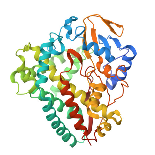
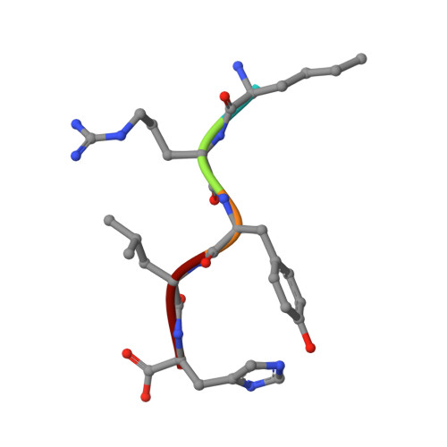
    RufO is a unique cytochrome P450 enzyme (CYP) involved in the biosynthesis of rufomycin, an antituberculosis cyclic peptide featuring an unusual nitrated tyrosine. Recent studies have clarified RufO's role in producing ribosomally synthesized and post-translationally modified peptides (RiPPs). Despite growing interest in nitrating enzymes and RiPP biosynthesis, the mechanism by which RufO recognizes and nitrates its pentapeptide substrate, MRYLH, remains poorly understood. In this study, we use a combination of spectroscopic, kinetic, and structural techniques to elucidate the molecular basis for peptide binding and heme-based nitration in RufO. Peptide binding is an endothermic process with a dissociation constant of 0.78 μM. Unlike most CYPs, RufO does not undergo the typical spin state conversion nor exhibit a significant increase in reduction potential upon substrate binding. The minimal perturbation to the heme center may lead to RufO's lack of specificity for redox partners. However, significant shifts in the vibrational frequencies of carbonyl complexes upon substrate binding indicate a more polar heme distal site that favors a nonlinear binding conformation of diatomic gas molecules. These distinctive features contrast with TxtE, the only other CYP known to catalyze aromatic nitration. A 1.51 Å resolution crystal structure reveals that substrate binding induces significant conformational changes in the distal pocket, particularly in the regions interacting with Arg-2 and His-5 of the MRYLH peptide. While Tyr-3 is positioned similarly to its counterpart in P450 Blt , a paralog that catalyzes peptide cross-linking, an extended hydrogen-bonding network constraining His-5 is unique to RufO and likely contributes to its distinct nitration activity. Furthermore, transient kinetic data suggest the sequential binding of O 2 followed by NO and characterize a ferric-superoxo intermediate essential for the nitration activity. This study provides valuable insights into the substrate specificity and catalytic mechanisms of CYPs involved in nitration reactions and RiPP biosynthesis.


  • Organizational Affiliation
    • Department of Chemistry, University of Georgia, Athens, Georgia 30602, United States.

Macromolecules
Find similar proteins by:  (by identity cutoff)  |  3D Structure
Entity ID: 1
MoleculeChains Sequence LengthOrganismDetailsImage
Cytochrome P450396Streptomyces atratusMutation(s): 0 
Gene Names: rufO
UniProt
Find proteins for A0A224AU14 (Streptomyces atratus)
Explore A0A224AU14 
Go to UniProtKB:  A0A224AU14
Entity Groups  
Sequence Clusters30% Identity50% Identity70% Identity90% Identity95% Identity100% Identity
UniProt GroupA0A224AU14
Sequence Annotations
Expand
  • Reference Sequence

Find similar proteins by:  Sequence   |   3D Structure  

Entity ID: 2
MoleculeChains Sequence LengthOrganismDetailsImage
NLE-ARG-TYR-LEU-HIS5Streptomyces atratusMutation(s): 0 
Sequence Annotations
Expand
  • Reference Sequence
Small Molecules
Ligands 4 Unique
IDChains Name / Formula / InChI Key2D Diagram3D Interactions
HEM (Subject of Investigation/LOI)
Query on HEM

Download Ideal Coordinates CCD File 
C [auth A]PROTOPORPHYRIN IX CONTAINING FE
C34 H32 Fe N4 O4
KABFMIBPWCXCRK-RGGAHWMASA-L
GOL
Query on GOL

Download Ideal Coordinates CCD File 
I [auth A],
J [auth A]
GLYCEROL
C3 H8 O3
PEDCQBHIVMGVHV-UHFFFAOYSA-N
ACT
Query on ACT

Download Ideal Coordinates CCD File 
E [auth A],
F [auth A],
G [auth A],
H [auth A]
ACETATE ION
C2 H3 O2
QTBSBXVTEAMEQO-UHFFFAOYSA-M
CL
Query on CL

Download Ideal Coordinates CCD File 
D [auth A]CHLORIDE ION
Cl
VEXZGXHMUGYJMC-UHFFFAOYSA-M
Modified Residues  1 Unique
IDChains TypeFormula2D DiagramParent
NLE
Query on NLE
B
L-PEPTIDE LINKINGC6 H13 N O2LEU
Experimental Data & Validation

Experimental Data

  • Method: X-RAY DIFFRACTION
  • Resolution: 1.51 Å
  • R-Value Free:  0.191 (Depositor), 0.193 (DCC) 
  • R-Value Work:  0.164 (Depositor), 0.164 (DCC) 
  • R-Value Observed: 0.165 (Depositor) 
Space Group: P 21 21 21
Unit Cell:
Length ( Å )Angle ( ˚ )
a = 56.258α = 90
b = 77.833β = 90
c = 89.178γ = 90
Software Package:
Software NamePurpose
PHENIXrefinement
SCALEPACKdata scaling
DENZOdata reduction
PHENIXphasing

Structure Validation

View Full Validation Report


Ligand Structure Quality Assessment 


Entry History & Funding Information

Deposition Data


Funding OrganizationLocationGrant Number
National Institutes of Health/National Institute of General Medical Sciences (NIH/NIGMS)United StatesR35GM147510

Revision History  (Full details and data files)

  • Version 1.0: 2025-06-18
    Type: Initial release
  • Version 1.1: 2025-07-09
    Changes: Database references