9DSF | pdb_00009dsf

Cyanide-ligated Bordetella pertussis globin coupled sensor regulatory domain S68A


Experimental Data Snapshot

  • Method: X-RAY DIFFRACTION
  • Resolution: 1.85 Å
  • R-Value Free: 
    0.239 (Depositor), 0.249 (DCC) 
  • R-Value Work: 
    0.207 (Depositor), 0.213 (DCC) 
  • R-Value Observed: 
    0.209 (Depositor) 

Starting Model: experimental
View more details

wwPDB Validation 3D Report Full Report

Validation slider image for 9DSF

Ligand Structure Quality Assessment 


This is version 1.1 of the entry. See complete history

Literature

Cooperative ligand binding in a bacterial heme-based oxygen sensor.

Hoque, N.J.Pope, S.R.Venkatakrishnan, V.Olori, D.O.Brady, N.A.Patterson, D.C.Anand, G.S.Liu, Y.Boal, A.K.Weinert, E.E.

(2025) J Biological Chem 302: 111025-111025

  • DOI: https://doi.org/10.1016/j.jbc.2025.111025
  • Primary Citation Related Structures: 
    9DSE, 9DSF

  • PubMed Abstract: 

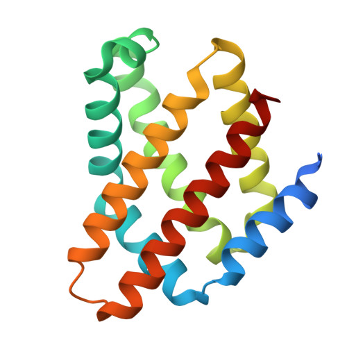
    Bacteria modulate essential phenotypes in response to external signals such as the availability of molecular oxygen (O 2 ). A class of direct O 2 -sensing heme proteins, globin coupled sensors, have been implicated in O 2 -dependent regulation of pathogenic phenotypes including biofilm formation, motility, and virulence. While cooperative O 2 binding is well known in both mammalian and prokaryotic hemoglobins, cooperative ligand binding previously has not been observed in bacterial sensor globins. This study explores the O 2 -dependent allosteric communication between globin domains in the globin-coupled sensor protein from Pectobacterium carotovorum (PccGCS) through equilibrium O 2 binding measurements, X-ray crystallography, resonance Raman spectroscopy, and hydrogen-deuterium exchange mass spectrometry. Based on these experiments, we propose a model of allosteric regulation of O 2 binding that is directed by subtle changes in distal heme pocket protein conformation and transduced through dynamics of helices at the dimer interface of the PccGCS sensor globin. Together this work identifies cooperative ligand binding in a family of bacterial heme proteins, which could allow the bacteria to more robustly respond to small changes in O 2 levels. Furthermore, this work highlights the importance of heme pocket residues in transducing the O 2 binding event within the dimer and suggests a pathway for signal transduction in dimeric myoglobin-like sensor proteins.


  • Organizational Affiliation
    • Department of Chemistry, Pennsylvania State University, University Park, PA 16802 USA.

Macromolecule Content 

  • Total Structure Weight: 59.46 kDa 
  • Atom Count: 3,898 
  • Modeled Residue Count: 467 
  • Deposited Residue Count: 510 
  • Unique protein chains: 1

Macromolecules

Find similar proteins by:|  3D Structure
Entity ID: 1
MoleculeChains  Sequence LengthOrganismDetailsImage
Diguanylate cyclase DosC
A, B, C
170Bordetella pertussis Tohama IMutation(s): 1 
Gene Names: BP3507
EC: 2.7.7.65
UniProt
Find proteins for Q7VTL8 (Bordetella pertussis (strain Tohama I / ATCC BAA-589 / NCTC 13251))
Explore Q7VTL8 
Go to UniProtKB:  Q7VTL8
Entity Groups
Sequence Clusters30% Identity50% Identity70% Identity90% Identity95% Identity100% Identity
UniProt GroupQ7VTL8
Sequence Annotations
Expand
Reference Sequence

Small Molecules

Ligands 3 Unique
IDChains Name / Formula / InChI Key2D Diagram3D Interactions
HEM
(Subject of Investigation/LOI)

Query on HEM


Download:Ideal Coordinates CCD File
D [auth A],
F [auth B],
I [auth C]
PROTOPORPHYRIN IX CONTAINING FE
C34 H32 Fe N4 O4
KABFMIBPWCXCRK-RGGAHWMASA-L
GOL

Query on GOL


Download:Ideal Coordinates CCD File
H [auth B]GLYCEROL
C3 H8 O3
PEDCQBHIVMGVHV-UHFFFAOYSA-N
CYN
(Subject of Investigation/LOI)

Query on CYN


Download:Ideal Coordinates CCD File
E [auth A],
G [auth B],
J [auth C]
CYANIDE ION
C N
XFXPMWWXUTWYJX-UHFFFAOYSA-N

Experimental Data & Validation

Experimental Data

  • Method: X-RAY DIFFRACTION
  • Resolution: 1.85 Å
  • R-Value Free:  0.239 (Depositor), 0.249 (DCC) 
  • R-Value Work:  0.207 (Depositor), 0.213 (DCC) 
  • R-Value Observed: 0.209 (Depositor) 
Space Group: C 1 2 1
Unit Cell:
Length ( Å )Angle ( ˚ )
a = 144.958α = 90
b = 49.702β = 110.56
c = 67.994γ = 90
Software Package:
Software NamePurpose
REFMACrefinement
HKL-2000data reduction
HKL-2000data scaling
PHASERphasing

Structure Validation

View Full Validation Report


Ligand Structure Quality Assessment 


Entry History 

& Funding Information

Deposition Data


Funding OrganizationLocationGrant Number
National Institutes of Health/National Institute of General Medical Sciences (NIH/NIGMS)United States--
National Science Foundation (NSF, United States)United States--

Revision History  (Full details and data files)

  • Version 1.0: 2025-12-24
    Type: Initial release
  • Version 1.1: 2026-01-14
    Changes: Database references