9DPZ | pdb_00009dpz

Potent inhibition of the protein arginine deiminases (PAD1-4) by targeting a Ca2+ dependent allosteric binding site


Experimental Data Snapshot

  • Method: X-RAY DIFFRACTION
  • Resolution: 2.41 Å
  • R-Value Free: 
    0.234 (Depositor), 0.234 (DCC) 
  • R-Value Work: 
    0.190 (Depositor), 0.191 (DCC) 
  • R-Value Observed: 
    0.192 (Depositor) 

Starting Model: experimental
View more details

wwPDB Validation 3D Report Full Report

Validation slider image for 9DPZ

Ligand Structure Quality Assessment 


This is version 1.0 of the entry. See complete history

Literature

Inhibiting peptidylarginine deiminases (PAD1-4) by targeting a Ca 2+ dependent allosteric binding site.

Dakin, L.A.Xing, L.Hall, J.Ding, W.Vajdos, F.F.Pelker, J.W.Ramsey, S.Balbo, P.Sahasrabudhe, P.V.Banker, M.E.Choi, W.Y.Wright, S.W.Chang, J.S.Curto, J.M.Davoren, J.E.Drozda, S.E.Fennell, K.F.Futatsugi, K.Kortum, S.Lee, K.L.Liu, S.Lovering, F.Nicki, J.A.Trujillo, J.I.Vincent, F.Schnute, M.E.

(2025) Nat Commun 16: 4579-4579

  • DOI: https://doi.org/10.1038/s41467-025-59919-4
  • Primary Citation Related Structures: 
    9DOL, 9DOP, 9DPZ

  • PubMed Abstract: 

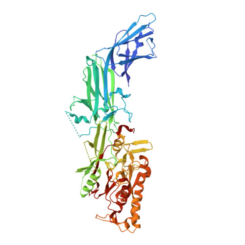
    Peptidylarginine deiminases (PAD1-4) are calcium dependent enzymes responsible for protein citrullination, a post-translational modification converting arginine residues to citrulline. Elevated levels of citrullinated proteins have been associated with rheumatoid arthritis, neurodegenerative diseases, and cancers. Though highly selective PAD4 inhibitors have been described, inhibitors to the broader family currently are limited to covalent substrate analogs. Herein, we describe an allosteric binding pocket common to PAD1-4 suitable for the identification of potent, non-covalent enzyme inhibitors. A ligand-based virtual screen is utilized to identify a PAD4 inhibitor for which surface plasmon resonance confirms target binding but non-competitively with a known PAD4 ligand. We further show through co-crystal structure analysis that the ligand binds PAD4 at an allosteric pocket resulting in stabilization of a catalytically inactive, calcium-deficient enzyme conformation. A ligand designed based on this site potently inhibits all four PAD isozymes and prevents protein citrullination in neutrophils with a broader protein repertoire than observed with a PAD4-selective inhibitor.


  • Organizational Affiliation
    • Medicine Design, Pfizer Inc, Cambridge, MA, USA.

Macromolecule Content 

  • Total Structure Weight: 75.95 kDa 
  • Atom Count: 4,627 
  • Modeled Residue Count: 596 
  • Deposited Residue Count: 671 
  • Unique protein chains: 1

Macromolecules

Find similar proteins by:|  3D Structure
Entity ID: 1
MoleculeChains  Sequence LengthOrganismDetailsImage
Protein-arginine deiminase type-4671Homo sapiensMutation(s): 3 
Gene Names: PADI4PAD4PADI5PDI5
EC: 3.5.3.15
UniProt & NIH Common Fund Data Resources
Find proteins for Q9UM07 (Homo sapiens)
Explore Q9UM07 
Go to UniProtKB:  Q9UM07
PHAROS:  Q9UM07
GTEx:  ENSG00000159339 
Entity Groups
Sequence Clusters30% Identity50% Identity70% Identity90% Identity95% Identity100% Identity
UniProt GroupQ9UM07
Sequence Annotations
Expand
Reference Sequence

Small Molecules

Ligands 2 Unique
IDChains Name / Formula / InChI Key2D Diagram3D Interactions
A1A9P
(Subject of Investigation/LOI)

Query on A1A9P


Download:Ideal Coordinates CCD File
B [auth A](2S)-2-amino-2-{1-[7-(4-bromophenyl)-5,6-dimethyl-7H-pyrrolo[2,3-d]pyrimidin-4-yl]piperidin-4-yl}-1-(pyrrolidin-1-yl)ethan-1-one
C25 H31 Br N6 O
TVTOXROCVZMGPW-QFIPXVFZSA-N
3YZ

Query on 3YZ


Download:Ideal Coordinates CCD File
C [auth A][(3S,4R)-3-amino-4-hydroxypiperidin-1-yl]{2-[1-(cyclopropylmethyl)-1H-pyrrolo[2,3-b]pyridin-2-yl]-7-methoxy-1-methyl-1H-benzimidazol-5-yl}methanone
C26 H30 N6 O3
SHBUAYSOPVZNDD-GHTZIAJQSA-N

Experimental Data & Validation

Experimental Data

  • Method: X-RAY DIFFRACTION
  • Resolution: 2.41 Å
  • R-Value Free:  0.234 (Depositor), 0.234 (DCC) 
  • R-Value Work:  0.190 (Depositor), 0.191 (DCC) 
  • R-Value Observed: 0.192 (Depositor) 
Space Group: C 1 2 1
Unit Cell:
Length ( Å )Angle ( ˚ )
a = 147.339α = 90
b = 60.421β = 123.357
c = 113.16γ = 90
Software Package:
Software NamePurpose
PHENIXrefinement
XDSdata reduction
autoPROCdata scaling
PHASERphasing

Structure Validation

View Full Validation Report


Ligand Structure Quality Assessment 


Entry History 

& Funding Information

Deposition Data

  • Released Date: 2025-05-28 
  • Deposition Author(s): Liu, S.

Funding OrganizationLocationGrant Number
Not funded--

Revision History  (Full details and data files)

  • Version 1.0: 2025-05-28
    Type: Initial release