9DPR | pdb_00009dpr

BMP-9 Wild-Type Dimer with Radiation Damage in Acidic pH

  • Classification: SIGNALING PROTEIN
  • Organism(s): Homo sapiens
  • Expression System: Homo sapiens
  • Mutation(s): Yes 

  • Deposited: 2024-09-23 Released: 2025-03-05 
  • Deposition Author(s): Schwartze, T.A., Hinck, A.P.
  • Funding Organization(s): National Institutes of Health/National Institute of General Medical Sciences (NIH/NIGMS), Department of Defense (DOD, United States)

Experimental Data Snapshot

  • Method: X-RAY DIFFRACTION
  • Resolution: 2.61 Å
  • R-Value Free: 
    0.266 (Depositor), 0.265 (DCC) 
  • R-Value Work: 
    0.212 (Depositor), 0.213 (DCC) 
  • R-Value Observed: 
    0.215 (Depositor) 

Starting Model: experimental
View more details

wwPDB Validation   3D Report Full Report


This is version 1.0 of the entry. See complete history


Literature

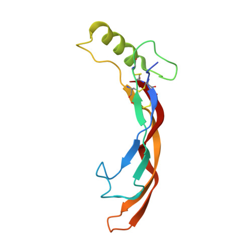
Molecular Basis of Interchain Disulfide Bond Formation in BMP-9 and BMP-10.

Schwartze, T.A.Morosky, S.A.Rosato, T.L.Henrickson, A.Lin, G.Hinck, C.S.Taylor, A.B.Olsen, S.K.Calero, G.Demeler, B.Roman, B.L.Hinck, A.P.

(2025) J Mol Biology 437: 168935-168935

  • DOI: https://doi.org/10.1016/j.jmb.2025.168935
  • Primary Citation of Related Structures:  
    9DPM, 9DPN, 9DPO, 9DPP, 9DPQ, 9DPR, 9DPS, 9DPT, 9DPU, 9DPV, 9DPW, 9DPX, 9DPY

  • PubMed Abstract: 

    BMP-9 and BMP-10 are TGF-β family signaling ligands naturally secreted into blood. They act on endothelial cells and are required for proper development and maintenance of the vasculature. In hereditary hemorrhagic telangiectasia, regulation is disrupted due to mutations in the BMP-9/10 pathway, namely in the type I receptor ALK1 or the co-receptor endoglin. It has been demonstrated that BMP-9/10 heterodimers are the most abundant signaling species in the blood, but it is unclear how they form. Unlike other ligands of the TGF-β family, BMP-9 and -10 are secreted as a mixture of disulfide-linked dimers and monomers in which the interchain cysteine (Cys-392) remains either unpaired or paired. Here, we show that the monomers are secreted in a cysteinylated form that crystallizes as a non-covalent dimer. Despite this, monomers do not self-associate at micromolar or lower concentrations and have reduced signaling potency compared to disulfide-linked dimers. We further show using protein crystallography that the interchain disulfide of the BMP-9 homodimer adopts a highly strained syn-periplanar conformation. Hence, geometric strain across the interchain disulfide is responsible for infrequent interchain disulfide bond formation, not the cysteinylation. Additionally, we show that interchain disulfide bond formation occurs less in BMP-9 than BMP-10 and these frequencies can be reversed by swapping residues near the interchain disulfide that form attractive interactions with the opposing protomer. Finally, we discuss the implications of these observations on BMP-9/10 heterodimer formation.


  • Organizational Affiliation

    Department of Structural Biology, School of Medicine, University of Pittsburgh, Pittsburgh, PA 15260, USA.


Macromolecules
Find similar proteins by:  (by identity cutoff)  |  3D Structure
Entity ID: 1
MoleculeChains Sequence LengthOrganismDetailsImage
Growth/differentiation factor 2110Homo sapiensMutation(s): 1 
Gene Names: GDF2BMP9
UniProt & NIH Common Fund Data Resources
Find proteins for Q9UK05 (Homo sapiens)
Explore Q9UK05 
Go to UniProtKB:  Q9UK05
PHAROS:  Q9UK05
GTEx:  ENSG00000263761 
Entity Groups  
Sequence Clusters30% Identity50% Identity70% Identity90% Identity95% Identity100% Identity
UniProt GroupQ9UK05
Sequence Annotations
Expand
  • Reference Sequence
Small Molecules
Ligands 3 Unique
IDChains Name / Formula / InChI Key2D Diagram3D Interactions
GOL
Query on GOL

Download Ideal Coordinates CCD File 
B [auth A]GLYCEROL
C3 H8 O3
PEDCQBHIVMGVHV-UHFFFAOYSA-N
CL
Query on CL

Download Ideal Coordinates CCD File 
C [auth A]
D [auth A]
E [auth A]
F [auth A]
G [auth A]
CHLORIDE ION
Cl
VEXZGXHMUGYJMC-UHFFFAOYSA-M
NA
Query on NA

Download Ideal Coordinates CCD File 
M [auth A],
N [auth A],
O [auth A]
SODIUM ION
Na
FKNQFGJONOIPTF-UHFFFAOYSA-N
Experimental Data & Validation

Experimental Data

  • Method: X-RAY DIFFRACTION
  • Resolution: 2.61 Å
  • R-Value Free:  0.266 (Depositor), 0.265 (DCC) 
  • R-Value Work:  0.212 (Depositor), 0.213 (DCC) 
  • R-Value Observed: 0.215 (Depositor) 
Space Group: I 41 2 2
Unit Cell:
Length ( Å )Angle ( ˚ )
a = 71.044α = 90
b = 71.044β = 90
c = 145.564γ = 90
Software Package:
Software NamePurpose
PHENIXrefinement
iMOSFLMdata reduction
Aimlessdata scaling
MOLREPphasing

Structure Validation

View Full Validation Report


Entry History & Funding Information

Deposition Data


Funding OrganizationLocationGrant Number
National Institutes of Health/National Institute of General Medical Sciences (NIH/NIGMS)United StatesGM58670
Department of Defense (DOD, United States)United StatesW81XWH-17-1-0429

Revision History  (Full details and data files)

  • Version 1.0: 2025-03-05
    Type: Initial release