9DNO | pdb_00009dno

Structure of UBR1-RFF complex


Experimental Data Snapshot

  • Method: X-RAY DIFFRACTION
  • Resolution: 1.33 Å
  • R-Value Free: 
    0.208 (Depositor), 0.208 (DCC) 
  • R-Value Work: 
    0.198 (Depositor), 0.201 (DCC) 
  • R-Value Observed: 
    0.199 (Depositor) 

Starting Model: experimental
View more details

wwPDB Validation   3D Report Full Report


This is version 1.1 of the entry. See complete history


Literature

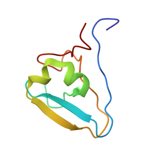
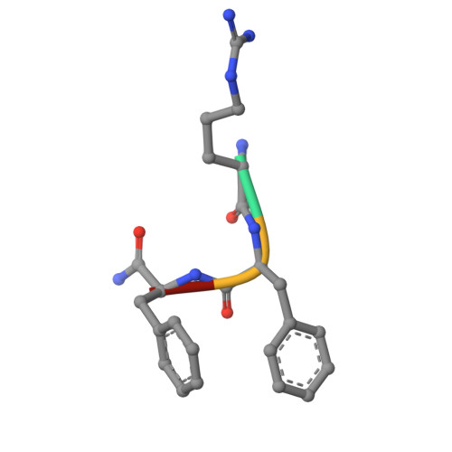
An alternative pocket for binding the N-degrons by the UBR1 and UBR2 ubiquitin E3 ligases.

Huang, S.T.Chen, D.H.Ren, T.Thomas, N.Wu, J.Sankaran, B.Jones, R.Taylor, S.Chen, Y.

(2025) Protein Sci 34: e70248-e70248

  • DOI: https://doi.org/10.1002/pro.70248
  • Primary Citation of Related Structures:  
    9DNO, 9DNP, 9DNQ, 9DNR, 9MUX

  • PubMed Abstract: 

    The UBR family of ubiquitin ligases binds to N-termini of their targets (known as N-degron) to induce their ubiquitination and degradation via a conserved domain known as UBR-box. UBR1 and UBR2 share the highest sequence homology among the family, and substantial structural studies were previously performed for substrate binding by the UBR-boxes of UBR1 and UBR2. Here, we describe a new pocket in the UBR-boxes of UBR1 and UBR2 for binding the second residues of N-degrons through determining five co-crystal structures of the UBR-boxes with various N-degron peptides. Together with binding affinities measured by fluorescence polarization, we show that the two highly homologous UBR-boxes can interact with the second residue of an N-degron differently. In addition, the UBR-boxes undergo different conformational changes when binding N-degrons. Furthermore, we demonstrate that the sidechain of the third amino acid of an N-degron has no contribution to binding the UBR-boxes. These findings represent a new conceptual advancement for the UBR E3 ligases and the new insights described here can be leveraged for developing their selective ligands for research and potential therapies.


  • Organizational Affiliation
    • Department of Chemistry and Biochemistry, University of California, San Diego, La Jolla, California, USA.

Macromolecules
Find similar proteins by:  (by identity cutoff)  |  3D Structure
Entity ID: 1
MoleculeChains Sequence LengthOrganismDetailsImage
E3 ubiquitin-protein ligase UBR1
A, B
77Homo sapiensMutation(s): 0 
Gene Names: UBR1
EC: 2.3.2.27
UniProt & NIH Common Fund Data Resources
Find proteins for Q8IWV7 (Homo sapiens)
Explore Q8IWV7 
Go to UniProtKB:  Q8IWV7
PHAROS:  Q8IWV7
GTEx:  ENSG00000159459 
Entity Groups  
Sequence Clusters30% Identity50% Identity70% Identity90% Identity95% Identity100% Identity
UniProt GroupQ8IWV7
Sequence Annotations
Expand
  • Reference Sequence

Find similar proteins by:  Sequence   |   3D Structure  

Entity ID: 2
MoleculeChains Sequence LengthOrganismDetailsImage
ARG-PHE-PHE-NH2C [auth D],
D [auth E]
4synthetic constructMutation(s): 0 
Sequence Annotations
Expand
  • Reference Sequence
Experimental Data & Validation

Experimental Data

  • Method: X-RAY DIFFRACTION
  • Resolution: 1.33 Å
  • R-Value Free:  0.208 (Depositor), 0.208 (DCC) 
  • R-Value Work:  0.198 (Depositor), 0.201 (DCC) 
  • R-Value Observed: 0.199 (Depositor) 
Space Group: P 1 21 1
Unit Cell:
Length ( Å )Angle ( ˚ )
a = 29.507α = 90
b = 48.952β = 106.85
c = 49.946γ = 90
Software Package:
Software NamePurpose
PHENIXrefinement
HKL-2000data reduction
HKL-2000data scaling
PHASERphasing

Structure Validation

View Full Validation Report


Entry History & Funding Information

Deposition Data


Funding OrganizationLocationGrant Number
Department of Defense (DOD, United States)United StatesHT9425-23-1-0560
Department of Defense (DOD, United States)United StatesPA220024P1
National Institutes of Health/National Cancer Institute (NIH/NCI)United StatesR01 CA265410
National Institutes of Health/National Cancer Institute (NIH/NCI)United StatesR01 CA212119

Revision History  (Full details and data files)

  • Version 1.0: 2025-08-13
    Type: Initial release
  • Version 1.1: 2026-02-25
    Changes: Database references