9DMB | pdb_00009dmb

Rhesus RHA10.01 Fab in complex with HIV-1 Env BG505 DS-SOSIP trimer


Experimental Data Snapshot

  • Method: ELECTRON MICROSCOPY
  • Resolution: 4.27 Å
  • Aggregation State: PARTICLE 
  • Reconstruction Method: SINGLE PARTICLE 

wwPDB Validation 3D Report Full Report

Validation slider image for 9DMB

This is version 1.1 of the entry. See complete history

Literature

Transient glycan shield reduction induces CD4-binding site broadly neutralizing antibodies in SHIV-infected macaques.

Morris, D.J.Gorman, J.Zhou, T.Lora, J.Connell, A.J.Li, H.Liu, W.Roark, R.S.Campion, M.S.Carey, J.W.Habib, R.Li, Y.Martella, C.L.Park, Y.Singh, A.Sowers, K.J.Teng, I.T.Wang, S.Chohan, N.Ding, W.Lauer, C.Lewis, E.Mason, R.D.Rando, J.M.Peyton, L.Schramm, C.A.Wagh, K.Korber, B.Seaman, M.S.Douek, D.C.Haynes, B.F.Kulp, D.W.Roederer, M.Hahn, B.H.Kwong, P.D.Shaw, G.M.

(2025) Cell Rep 44: 115848-115848

  • DOI: https://doi.org/10.1016/j.celrep.2025.115848
  • Primary Citation Related Structures: 
    9DMB

  • PubMed Abstract: 

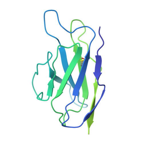
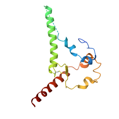
    Broadly neutralizing antibodies (bNAbs) targeting the HIV-1 CD4-binding site (CD4bs) occur infrequently in macaques and humans and have not been reproducibly elicited in any outbred animal model. To address this challenge, we first isolated RHA10, an infection-induced rhesus bNAb with 51% breadth. The cryoelectron microscopy (cryo-EM) structure of RHA10 with the HIV-1 envelope (Env) resembled prototypic human CD4bs bNAbs with CDR-H3-dominated binding. Env-antibody co-evolution revealed transient elimination of two Env CD4bs-proximal glycans near the time of RHA10-lineage initiation, and these glycan-deficient Envs bound preferentially to early RHA10 intermediates, suggesting that glycan deletions in infecting SHIVs could induce CD4bs bNAbs. To test this hypothesis, we constructed SHIV.CH505 variants with CD4bs-proximal glycan deletions. Infection of 11 macaques resulted in accelerated CD4bs bNAb responses in 9 compared with 1 of 115 control macaques. Glycan hole-based immunofocusing coupled to Env-Ab co-evolution can consistently induce broad CD4bs responses in macaques and serve as a model for HIV vaccine design.


  • Organizational Affiliation
    • Departments of Medicine and Microbiology, Perelman School of Medicine, University of Pennsylvania, Philadelphia, PA, USA.

Macromolecule Content 

  • Total Structure Weight: 391.02 kDa 
  • Atom Count: 21,021 
  • Modeled Residue Count: 2,448 
  • Deposited Residue Count: 3,285 
  • Unique protein chains: 4

Macromolecules

Find similar proteins by:|  3D Structure
Entity ID: 1
MoleculeChains  Sequence LengthOrganismDetailsImage
RHA10.01 Light chainA,
D [auth B],
G [auth D]
219Macaca mulattaMutation(s): 0 
Find similar proteins by:|  3D Structure
Entity ID: 2
MoleculeChains  Sequence LengthOrganismDetailsImage
RHA10.01 Heavy chainB [auth C],
E [auth G],
H
243Macaca mulattaMutation(s): 0 
Find similar proteins by:|  3D Structure
Entity ID: 3
MoleculeChains  Sequence LengthOrganismDetailsImage
BG505 DS-SOSIP glycoprotein gp41C [auth E],
F [auth I],
I [auth J]
153Human immunodeficiency virus 1Mutation(s): 2 
Gene Names: env
UniProt
Find proteins for A0ABF7PQ88 (Human immunodeficiency virus type 1)
Explore A0ABF7PQ88 
Go to UniProtKB:  A0ABF7PQ88
Entity Groups
Sequence Clusters30% Identity50% Identity70% Identity90% Identity95% Identity100% Identity
UniProt GroupA0ABF7PQ88
Glycosylation
Glycosylation Sites: 2
Sequence Annotations
Expand
Reference Sequence
Find similar proteins by:|  3D Structure
Entity ID: 4
MoleculeChains  Sequence LengthOrganismDetailsImage
Envelope glycoprotein gp120J [auth L],
K [auth F],
L [auth K]
480Human immunodeficiency virus 1Mutation(s): 0 
Gene Names: env
UniProt
Find proteins for Q2N0S6 (Human immunodeficiency virus type 1)
Explore Q2N0S6 
Go to UniProtKB:  Q2N0S6
Entity Groups
Sequence Clusters30% Identity50% Identity70% Identity90% Identity95% Identity100% Identity
UniProt GroupQ2N0S6
Glycosylation
Glycosylation Sites: 19
Sequence Annotations
Expand
Reference Sequence

Oligosaccharides

Help  
Entity ID: 5
MoleculeChains Length2D Diagram GlycosylationD Interactions
2-acetamido-2-deoxy-beta-D-glucopyranose-(1-4)-2-acetamido-2-deoxy-beta-D-glucopyranose
AA [auth a],
GA [auth g],
JA [auth j],
KA [auth k],
M,
AA [auth a],
GA [auth g],
JA [auth j],
KA [auth k],
M,
MA [auth m],
O,
SA [auth s],
U,
VA [auth v],
X,
Y
2N-Glycosylation
Glycosylation Resources
GlyTouCan: G42666HT
GlyCosmos: G42666HT
GlyGen: G42666HT
Entity ID: 6
MoleculeChains Length2D Diagram GlycosylationD Interactions
alpha-D-mannopyranose-(1-3)-[alpha-D-mannopyranose-(1-6)]beta-D-mannopyranose-(1-4)-2-acetamido-2-deoxy-beta-D-glucopyranose-(1-4)-2-acetamido-2-deoxy-beta-D-glucopyranose
CA [auth c],
EA [auth e],
LA [auth l],
N,
OA [auth o],
CA [auth c],
EA [auth e],
LA [auth l],
N,
OA [auth o],
Q,
QA [auth q],
S,
Z
5N-Glycosylation
Glycosylation Resources
GlyTouCan: G22768VO
GlyCosmos: G22768VO
GlyGen: G22768VO
Entity ID: 7
MoleculeChains Length2D Diagram GlycosylationD Interactions
beta-D-mannopyranose-(1-4)-2-acetamido-2-deoxy-beta-D-glucopyranose-(1-4)-2-acetamido-2-deoxy-beta-D-glucopyranose
BA [auth b],
DA [auth d],
FA [auth f],
HA [auth h],
IA [auth i],
BA [auth b],
DA [auth d],
FA [auth f],
HA [auth h],
IA [auth i],
NA [auth n],
P,
PA [auth p],
R,
RA [auth r],
T,
TA [auth t],
UA [auth u],
V,
W
3N-Glycosylation
Glycosylation Resources
GlyTouCan: G15407YE
GlyCosmos: G15407YE
GlyGen: G15407YE

Small Molecules

Ligands 1 Unique
IDChains Name / Formula / InChI Key2D Diagram3D Interactions
NAG

Query on NAG


Download:Ideal Coordinates CCD File
AB [auth J]
BB [auth J]
CB [auth L]
DB [auth L]
EB [auth L]
AB [auth J],
BB [auth J],
CB [auth L],
DB [auth L],
EB [auth L],
FB [auth L],
GB [auth L],
HB [auth L],
IB [auth L],
JB [auth F],
KB [auth F],
LB [auth F],
MB [auth F],
NB [auth F],
OB [auth F],
PB [auth F],
QB [auth K],
RB [auth K],
SB [auth K],
TB [auth K],
UB [auth K],
VB [auth K],
WA [auth E],
WB [auth K],
XA [auth E],
YA [auth I],
ZA [auth I]
2-acetamido-2-deoxy-beta-D-glucopyranose
C8 H15 N O6
OVRNDRQMDRJTHS-FMDGEEDCSA-N

Experimental Data & Validation

Experimental Data

  • Method: ELECTRON MICROSCOPY
  • Resolution: 4.27 Å
  • Aggregation State: PARTICLE 
  • Reconstruction Method: SINGLE PARTICLE 
EM Software:
TaskSoftware PackageVersion
RECONSTRUCTIONcryoSPARC

Structure Validation

View Full Validation Report


Entry History 

& Funding Information

Deposition Data


Funding OrganizationLocationGrant Number
National Institutes of Health/National Institute of General Medical Sciences (NIH/NIGMS)United StatesGM103310
Other privateUnited StatesSimons Foundation (SF349247)

Revision History  (Full details and data files)

  • Version 1.0: 2025-06-18
    Type: Initial release
  • Version 1.1: 2025-06-25
    Changes: Data collection, Database references