9DIF | pdb_00009dif

CBASS Pseudomonas syringae Cap5 tetramer with DNA duplex and 3'2'-c-GAMP cyclic dinucleotide ligand


Experimental Data Snapshot

  • Method: X-RAY DIFFRACTION
  • Resolution: 1.67 Å
  • R-Value Free: 
    0.211 (Depositor), 0.211 (DCC) 
  • R-Value Work: 
    0.170 (Depositor), 0.170 (DCC) 
  • R-Value Observed: 
    0.172 (Depositor) 

Starting Model: experimental
View more details

wwPDB Validation 3D Report Full Report

Validation slider image for 9DIF

Ligand Structure Quality Assessment 


This is version 1.0 of the entry. See complete history

Literature

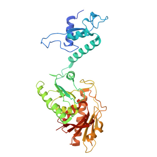
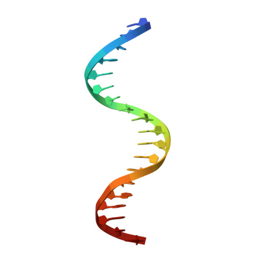
Mechanism of DNA degradation by CBASS Cap5 endonuclease immune effector.

Rechkoblit, O.Sciaky, D.Ni, M.Li, Y.Kottur, J.Fang, G.Aggarwal, A.K.

(2025) Nat Commun 16: 5243-5243

  • DOI: https://doi.org/10.1038/s41467-025-60484-z
  • Primary Citation Related Structures: 
    9DIF, 9DIH, 9NLG

  • PubMed Abstract: 

    Bacterial CBASS immune defense systems commonly kill virally infected cells by degrading genomic DNA in a form of cell suicide or abortive infection. We present a high-resolution structure of the CBASS effector Cap5, activated by a cyclic nucleotide, in the act of digesting DNA via tetrameric HNH endonuclease domains. Two HNH domains are in a catalytically active state for cleavage of the DNA strands, whereas the other two HNH domains are in a topologically distinct catalytically inactive state for simply DNA binding. The four HNH domains track one face of the DNA and mark an enzyme that acts as a stand-alone non-specific nuclease. We also show that chromosomally encoded CBASS Cap5 can be extrinsically activated by a cyclic nucleotide, as a step towards potential antibiotics.


  • Organizational Affiliation
    • Department of Pharmacological Sciences, Icahn School of Medicine at Mount Sinai, New York, NY, USA. olga.rechkoblit@mssm.edu.

Macromolecule Content 

  • Total Structure Weight: 92.7 kDa 
  • Atom Count: 7,212 
  • Modeled Residue Count: 750 
  • Deposited Residue Count: 795 
  • Unique protein chains: 1
  • Unique nucleic acid chains: 1

Macromolecules


Find similar proteins by:|  3D Structure
Entity ID: 1
MoleculeChains  Sequence LengthOrganismDetailsImage
HNH endonuclease
A, B
388Pseudomonas syringaeMutation(s): 1 
UniProt
Find proteins for A0A2P0QGK5 (Pseudomonas syringae pv. actinidiae)
Explore A0A2P0QGK5 
Go to UniProtKB:  A0A2P0QGK5
Entity Groups
Sequence Clusters30% Identity50% Identity70% Identity90% Identity95% Identity100% Identity
UniProt GroupA0A2P0QGK5
Sequence Annotations
Expand
Reference Sequence
Find similar nucleic acids by:  Sequence
Entity ID: 2
MoleculeChains LengthOrganismImage
DNA (5'-D(*TP*TP*GP*CP*TP*CP*TP*CP*TP*TP*AP*AP*GP*AP*GP*AP*GP*CP*A)-3')C [auth D]19synthetic construct
Sequence Annotations
Expand
Reference Sequence

Experimental Data & Validation

Experimental Data

  • Method: X-RAY DIFFRACTION
  • Resolution: 1.67 Å
  • R-Value Free:  0.211 (Depositor), 0.211 (DCC) 
  • R-Value Work:  0.170 (Depositor), 0.170 (DCC) 
  • R-Value Observed: 0.172 (Depositor) 
Space Group: C 2 2 21
Unit Cell:
Length ( Å )Angle ( ˚ )
a = 105.021α = 90
b = 194.171β = 90
c = 118.626γ = 90
Software Package:
Software NamePurpose
PHENIXrefinement
XDSdata reduction
Aimlessdata scaling
PHASERphasing

Structure Validation

View Full Validation Report


Ligand Structure Quality Assessment 


Entry History 

& Funding Information

Deposition Data


Funding OrganizationLocationGrant Number
National Institutes of Health/National Institute of General Medical Sciences (NIH/NIGMS)United StatesR35-GM131780

Revision History  (Full details and data files)

  • Version 1.0: 2025-06-18
    Type: Initial release