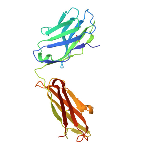
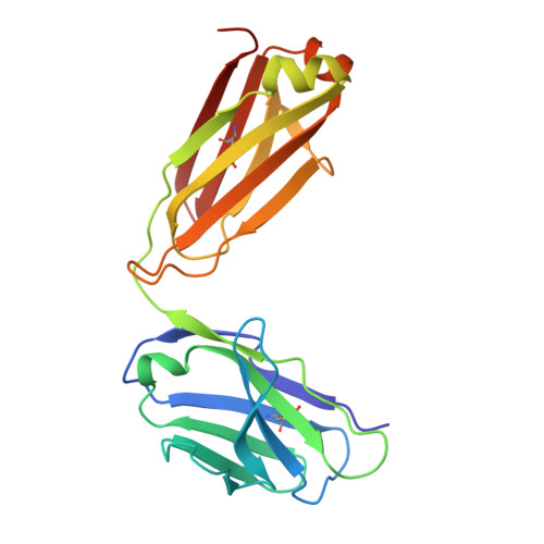
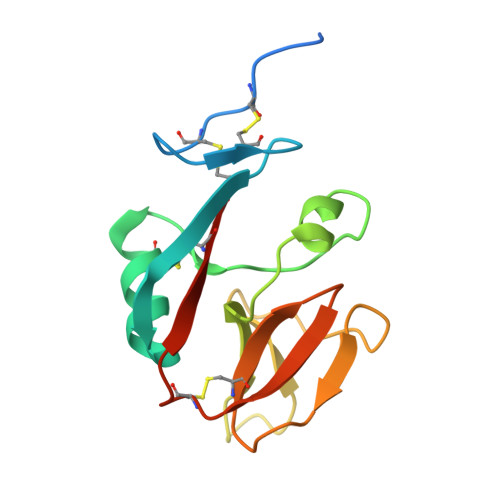
9DH2

Structure of Fab in complex with NKG2D extracellular domain


Experimental Data Snapshot

  • Method: X-RAY DIFFRACTION
  • Resolution: 2.98 Å
  • R-Value Free: 0.243 
  • R-Value Work: 0.197 
  • R-Value Observed: 0.199 

Starting Models: experimental
View more details

wwPDB Validation   3D Report Full Report


This is version 1.2 of the entry. See complete history


Literature

Agonistic anti-NKG2D antibody structure reveals unique stoichiometry and epitope compared to natural ligands.

Fallon, D.Huang, C.S.Ma, J.Morgan, C.Zhou, Z.S.

(2024) MAbs 16: 2433121-2433121

  • DOI: https://doi.org/10.1080/19420862.2024.2433121
  • Primary Citation of Related Structures:  
    9DH2

  • PubMed Abstract: 

    Natural killer (NK) cells are effector cells of the innate immune system that distinguish between healthy and abnormal cells through activating and inhibitory receptor signaling. NKG2D, a homodimeric activating receptor expressed on NK cells, recognizes a diverse class of stress ligands expressed by cells experiencing infection, malignant transformation, chronic inflammation, and other cellular stresses. Despite the variety of NKG2D ligands, they all bind the receptor asymmetrically in a 1:1 ligand to homodimeric NKG2D stoichiometry. In contrast, as we report herein, the agonistic antibody 2D3 binds NKG2D with a 2:1 stoichiometry of its antigen binding fragments to homodimeric NKG2D and a largely distinct epitope. This binding interaction, as compared to NKG2D natural ligands, suggests there may be unique mechanisms to engage this receptor while offering possible benefits when incorporated into an IgG-based therapeutic.


  • Organizational Affiliation

    Dragonfly Therapeutics, Inc., Waltham, MA, USA.


Macromolecules
Find similar proteins by:  (by identity cutoff)  |  3D Structure
Entity ID: 1
MoleculeChains Sequence LengthOrganismDetailsImage
Fab heavy chainA,
C [auth G],
E [auth K],
H [auth P]
220Homo sapiensMutation(s): 0 
Entity Groups  
Sequence Clusters30% Identity50% Identity70% Identity90% Identity95% Identity100% Identity
Sequence Annotations
Expand
  • Reference Sequence
Find similar proteins by:  (by identity cutoff)  |  3D Structure
Entity ID: 2
MoleculeChains Sequence LengthOrganismDetailsImage
Fab light chainB [auth D],
D [auth H],
F [auth L],
I [auth Q]
212Homo sapiensMutation(s): 0 
Entity Groups  
Sequence Clusters30% Identity50% Identity70% Identity90% Identity95% Identity100% Identity
Sequence Annotations
Expand
  • Reference Sequence
Find similar proteins by:  (by identity cutoff)  |  3D Structure
Entity ID: 3
MoleculeChains Sequence LengthOrganismDetailsImage
NKG2-D type II integral membrane proteinG [auth M],
J [auth R],
K [auth S],
L [auth T]
137Homo sapiensMutation(s): 0 
Gene Names: KLRK1D12S2489ENKG2D
UniProt & NIH Common Fund Data Resources
Find proteins for P26718 (Homo sapiens)
Explore P26718 
Go to UniProtKB:  P26718
PHAROS:  P26718
GTEx:  ENSG00000213809 
Entity Groups  
Sequence Clusters30% Identity50% Identity70% Identity90% Identity95% Identity100% Identity
UniProt GroupP26718
Sequence Annotations
Expand
  • Reference Sequence
Experimental Data & Validation

Experimental Data

  • Method: X-RAY DIFFRACTION
  • Resolution: 2.98 Å
  • R-Value Free: 0.243 
  • R-Value Work: 0.197 
  • R-Value Observed: 0.199 
  • Space Group: P 1 21 1
Unit Cell:
Length ( Å )Angle ( ˚ )
a = 47.062α = 90
b = 224.433β = 91.22
c = 136.297γ = 90
Software Package:
Software NamePurpose
REFMACrefinement
autoPROCdata reduction
autoPROCdata scaling
PHASERphasing

Structure Validation

View Full Validation Report


Entry History & Funding Information

Deposition Data


Funding OrganizationLocationGrant Number
Not funded--

Revision History  (Full details and data files)

  • Version 1.0: 2024-12-04
    Type: Initial release
  • Version 1.1: 2024-12-11
    Changes: Database references
  • Version 1.2: 2024-12-18
    Changes: Derived calculations