9D84 | pdb_00009d84

Shigella flexneri bacteriophage B2 Gp48 and Gp49


Experimental Data Snapshot

  • Method: ELECTRON MICROSCOPY
  • Resolution: 2.40 Å
  • Aggregation State: PARTICLE 
  • Reconstruction Method: SINGLE PARTICLE 

wwPDB Validation   3D Report Full Report


This is version 1.0 of the entry. See complete history


Literature

Moo19 and B2: Structures of Schitoviridae podophages with T = 9 geometry and tailspikes with esterase activity.

Subramanian, S.Bergland Drarvik, S.M.Tinney, K.R.Doore, S.M.Parent, K.N.

(2024) Sci Adv 10: eadt0022-eadt0022

  • DOI: https://doi.org/10.1126/sciadv.adt0022
  • Primary Citation of Related Structures:  
    9D7Z, 9D80, 9D81, 9D82, 9D83, 9D84

  • PubMed Abstract: 

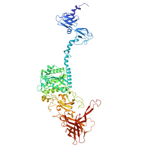
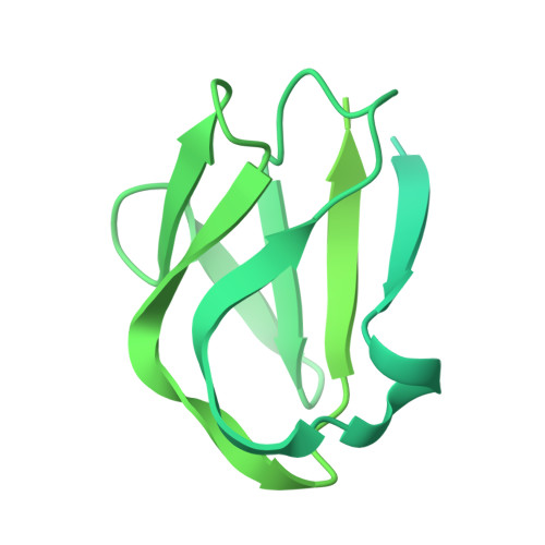
    Podophages are, by far, the least well studied of all the bacteriophages. Despite being classified together due to their short, noncontractile tails, there is a huge amount of diversity among members of this group. Of the podophages, the N4-like Schitoviridae family is the least well studied structurally and is quite divergent from well-characterized podophages such as T7 and P22. In this work, we isolate and fully characterize two members of the Schitoviridae family by cryo-electron microscopy, genetics, and biochemistry. We describe the capsid features of Moo19 and B2, including a decoration protein. In addition, we have fully modeled the tail machinery for both phages and identify proteins with esterase activity. Genetic knockouts of the host reveal factors specific for host attachment including key modifications to the O-antigen on the lipopolysaccharide. Moo19 and B2 are both Schitoviridae members, yet some distinct differences in the genome and structure place them into distinct clades.


  • Organizational Affiliation
    • Department of Biochemistry and Molecular Biology, Michigan State University, East Lansing, MI 48824, USA.

Macromolecules
Find similar proteins by:  (by identity cutoff)  |  3D Structure
Entity ID: 1
MoleculeChains Sequence LengthOrganismDetailsImage
Gp48777Shigella phage B2Mutation(s): 0 
Entity Groups  
Sequence Clusters30% Identity50% Identity70% Identity90% Identity95% Identity100% Identity
Sequence Annotations
Expand
  • Reference Sequence
Find similar proteins by:  (by identity cutoff)  |  3D Structure
Entity ID: 2
MoleculeChains Sequence LengthOrganismDetailsImage
Gp49410Shigella phage B2Mutation(s): 0 
Entity Groups  
Sequence Clusters30% Identity50% Identity70% Identity90% Identity95% Identity100% Identity
Sequence Annotations
Expand
  • Reference Sequence
Experimental Data & Validation

Experimental Data

  • Method: ELECTRON MICROSCOPY
  • Resolution: 2.40 Å
  • Aggregation State: PARTICLE 
  • Reconstruction Method: SINGLE PARTICLE 
EM Software:
TaskSoftware PackageVersion
MODEL REFINEMENTPHENIX1.20.1_4487:

Structure Validation

View Full Validation Report


Entry History & Funding Information

Deposition Data


Funding OrganizationLocationGrant Number
National Institutes of Health/National Institute of General Medical Sciences (NIH/NIGMS)United StatesGM110185
National Institutes of Health/National Institute of General Medical Sciences (NIH/NIGMS)United StatesGM116789
National Institutes of Health/National Institute of General Medical Sciences (NIH/NIGMS)United StatesGM140803
National Science Foundation (NSF, United States)United States1750125

Revision History  (Full details and data files)

  • Version 1.0: 2025-01-15
    Type: Initial release