9D4Y | pdb_00009d4y

Structure of PAK1 in complex with compound 31


Experimental Data Snapshot

  • Method: X-RAY DIFFRACTION
  • Resolution: 1.85 Å
  • R-Value Free: 
    0.227 (Depositor), 0.228 (DCC) 
  • R-Value Work: 
    0.188 (Depositor), 0.196 (DCC) 

Starting Model: experimental
View more details

wwPDB Validation 3D Report Full Report

Validation slider image for 9D4Y

Ligand Structure Quality Assessment 


This is version 1.2 of the entry. See complete history

Literature

Identification of a p21-activated kinase 1 (PAK1) inhibitor with 10-fold selectivity against PAK2.

Johns, D.M.Olejniczak, J.Babbar, A.Boone, C.D.Cakici, O.Cheng, M.Cheng, Q.Q.Dementiev, A.Eick, M.Ferdyan, N.Fontano, E.Forman, A.Kozlowski, R.Lee, S.W.Mehta, S.Mowery, K.Murray, B.Nguyen, V.Olland, A.Phan, K.B.Rivera, L.Sabat, M.Sprengeler, P.Srinivasan, K.Sun, Z.Suto, R.K.Wilkinson, T.Wang, C.Yu, N.Xu, M.Goel, V.Hirst, G.Reich, S.

(2025) Bioorg Med Chem Lett 127: 130307-130307

  • DOI: https://doi.org/10.1016/j.bmcl.2025.130307
  • Primary Citation Related Structures: 
    9D4V, 9D4W, 9D4X, 9D4Y, 9D50, 9D51, 9D52, 9D53

  • PubMed Abstract: 

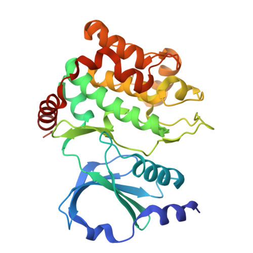
    The p21-activated kinases (PAKs) are noted for their role in cytoskeletal organization, cellular morphogenesis, and pro-survival signaling. PAK1 is of particular interest due to its role in tumorigenesis, being amplified in multiple cancers (the most prevalent being breast, ovarian, and melanoma cancers). PAK2 is closely related to PAK1 in structure but is associated with cardiotoxicity. A structure-based design effort targeting a PAK1 (over PAK2) selective small molecule inhibitor is detailed herein. We report here the first crystal structure of PAK2 and use this crystal structure to design a PAK1 inhibitor with ten-fold selectivity over PAK2.


  • Organizational Affiliation
    • Turning Point Therapeutics, Inc. (a wholly owned subsidiary of Bristol Myers Squibb Company), 10628 Science Center Drive, Suite 200, San Diego, CA 92121, United States. Electronic address: deidre123@gmail.com.

Macromolecule Content 

  • Total Structure Weight: 67.48 kDa 
  • Atom Count: 4,582 
  • Modeled Residue Count: 571 
  • Deposited Residue Count: 594 
  • Unique protein chains: 1

Macromolecules

Find similar proteins by:|  3D Structure
Entity ID: 1
MoleculeChains  Sequence LengthOrganismDetailsImage
Serine/threonine-protein kinase PAK 1
A, B
297Homo sapiensMutation(s): 2 
Gene Names: PAK1
EC: 2.7.11.1
UniProt & NIH Common Fund Data Resources
Find proteins for Q13153 (Homo sapiens)
Explore Q13153 
Go to UniProtKB:  Q13153
PHAROS:  Q13153
GTEx:  ENSG00000149269 
Entity Groups
Sequence Clusters30% Identity50% Identity70% Identity90% Identity95% Identity100% Identity
UniProt GroupQ13153
Sequence Annotations
Expand
Reference Sequence

Small Molecules

Ligands 1 Unique
IDChains Name / Formula / InChI Key2D Diagram3D Interactions
A1A2S
(Subject of Investigation/LOI)

Query on A1A2S


Download:Ideal Coordinates CCD File
C [auth A],
D [auth B]
N~2~-{[(1R,3R,4S)-4-amino-3-(3-chlorophenyl)cyclohexyl]methyl}-N~4~-(5-cyclopropyl-1,3-thiazol-2-yl)pyrimidine-2,4-diamine
C23 H27 Cl N6 S
RPZYQSABIKMHOM-ZMYBRWDISA-N

Experimental Data & Validation

Experimental Data

  • Method: X-RAY DIFFRACTION
  • Resolution: 1.85 Å
  • R-Value Free:  0.227 (Depositor), 0.228 (DCC) 
  • R-Value Work:  0.188 (Depositor), 0.196 (DCC) 
Space Group: P 1 21 1
Unit Cell:
Length ( Å )Angle ( ˚ )
a = 63.306α = 90
b = 80.946β = 106.61
c = 65.883γ = 90
Software Package:
Software NamePurpose
REFMACrefinement
XDSdata reduction
XDSdata scaling
PHASERphasing

Structure Validation

View Full Validation Report


Ligand Structure Quality Assessment 


Entry History 

& Funding Information

Deposition Data


Funding OrganizationLocationGrant Number
Other private--

Revision History  (Full details and data files)

  • Version 1.0: 2025-04-02
    Type: Initial release
  • Version 1.1: 2025-06-25
    Changes: Database references
  • Version 1.2: 2025-07-16
    Changes: Database references