9D3E | pdb_00009d3e

Cryo-EM structure of CCR6 bound by SQA1 and OXM2


Experimental Data Snapshot

  • Method: ELECTRON MICROSCOPY
  • Resolution: 3.02 Å
  • Aggregation State: PARTICLE 
  • Reconstruction Method: SINGLE PARTICLE 

wwPDB Validation 3D Report Full Report

Validation slider image for 9D3E

This is version 1.1 of the entry. See complete history

Literature

Structural basis for CCR6 modulation by allosteric antagonists.

Wasilko, D.J.Gerstenberger, B.S.Farley, K.A.Li, W.Alley, J.Schnute, M.E.Unwalla, R.J.Victorino, J.Crouse, K.K.Ding, R.Sahasrabudhe, P.V.Vincent, F.Frisbie, R.K.Dermenci, A.Flick, A.Choi, C.Chinigo, G.Mousseau, J.J.Trujillo, J.I.Nuhant, P.Mondal, P.Lombardo, V.Lamb, D.Hogan, B.J.Minhas, G.S.Segala, E.Oswald, C.Windsor, I.W.Han, S.Rappas, M.Cooke, R.M.Calabrese, M.F.Berstein, G.Thorarensen, A.Wu, H.

(2024) Nat Commun 15: 7574-7574

  • DOI: https://doi.org/10.1038/s41467-024-52045-7
  • Primary Citation Related Structures: 
    9D3E, 9D3G

  • PubMed Abstract: 

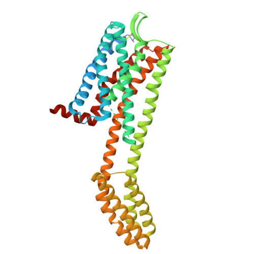
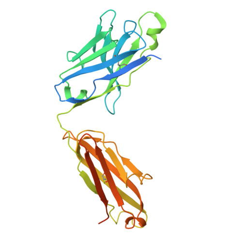
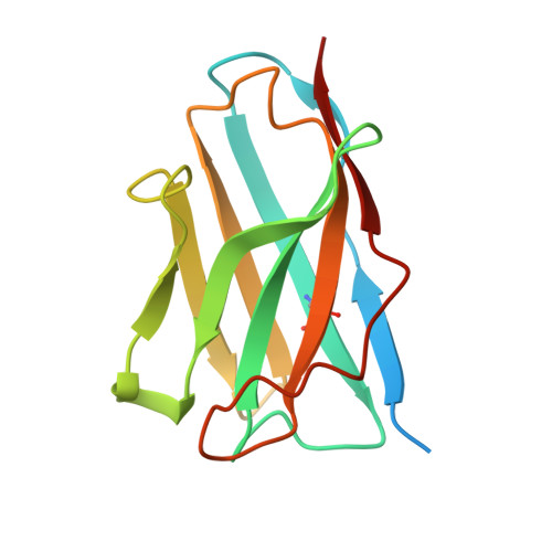
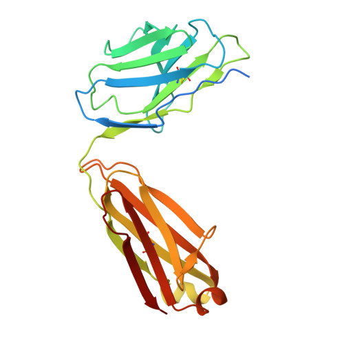
    The CC chemokine receptor 6 (CCR6) is a potential target for chronic inflammatory diseases. Previously, we reported an active CCR6 structure in complex with its cognate chemokine CCL20, revealing the molecular basis of CCR6 activation. Here, we present two inactive CCR6 structures in ternary complexes with different allosteric antagonists, CCR6/SQA1/OXM1 and CCR6/SQA1/OXM2. The oxomorpholine analogues, OXM1 and OXM2 are highly selective CCR6 antagonists which bind to an extracellular pocket and disrupt the receptor activation network. An energetically favoured U-shaped conformation in solution that resembles the bound form is observed for the active analogues. SQA1 is a squaramide derivative with close-in analogues reported as antagonists of chemokine receptors including CCR6. SQA1 binds to an intracellular pocket which overlaps with the G protein site, stabilizing a closed pocket that is a hallmark of inactive GPCRs. Minimal communication between the two allosteric pockets is observed, in contrast to the prevalent allosteric cooperativity model of GPCRs. This work highlights the versatility of GPCR antagonism by small molecules, complementing previous knowledge of CCR6 activation, and sheds light on drug discovery targeting CCR6.


  • Organizational Affiliation
    • Discovery Sciences, Medicine Design, Pfizer Inc., Groton, CT, USA.

Macromolecule Content 

  • Total Structure Weight: 125.78 kDa 
  • Atom Count: 7,173 
  • Modeled Residue Count: 917 
  • Deposited Residue Count: 1,123 
  • Unique protein chains: 4

Macromolecules

Find similar proteins by:|  3D Structure
Entity ID: 1
MoleculeChains  Sequence LengthOrganismDetailsImage
Human CCR6480Homo sapiensMutation(s): 0 
Gene Names: CCR6
Find similar proteins by:|  3D Structure
Entity ID: 2
MoleculeChains  Sequence LengthOrganismDetailsImage
anti-BRIL Fab Heavy chainB [auth H]269Homo sapiensMutation(s): 0 
Find similar proteins by:|  3D Structure
Entity ID: 3
MoleculeChains  Sequence LengthOrganismDetailsImage
anti-BRIL Fab NanobodyC [auth K]141synthetic constructMutation(s): 0 
Find similar proteins by:|  3D Structure
Entity ID: 4
MoleculeChains  Sequence LengthOrganismDetailsImage
anti-BRIL Fab Light chainD [auth L]233Homo sapiensMutation(s): 0 

Small Molecules

Ligands 3 Unique
IDChains Name / Formula / InChI Key2D Diagram3D Interactions
EBX
(Subject of Investigation/LOI)

Query on EBX


Download:Ideal Coordinates CCD File
F [auth A]4-[[3,4-bis(oxidanylidene)-2-[[(1~{R})-1-(4-propan-2-ylfuran-2-yl)propyl]amino]cyclobuten-1-yl]amino]-~{N},~{N}-dimethyl-3-oxidanyl-pyridine-2-carboxamide
C22 H26 N4 O5
JSZQMZJVSCVDMA-CYBMUJFWSA-N
A1A1W
(Subject of Investigation/LOI)

Query on A1A1W


Download:Ideal Coordinates CCD File
G [auth A]N-{[(2R)-4-(bicyclo[1.1.1]pentan-1-yl)-5-oxomorpholin-2-yl]methyl}-1-[4-(trifluoromethyl)phenyl]cyclopropane-1-carboxamide
C21 H23 F3 N2 O3
MGXLQVNOTOHKMK-VVFCZOMOSA-N
CLR

Query on CLR


Download:Ideal Coordinates CCD File
E [auth A]CHOLESTEROL
C27 H46 O
HVYWMOMLDIMFJA-DPAQBDIFSA-N

Experimental Data & Validation

Experimental Data

  • Method: ELECTRON MICROSCOPY
  • Resolution: 3.02 Å
  • Aggregation State: PARTICLE 
  • Reconstruction Method: SINGLE PARTICLE 
EM Software:
TaskSoftware PackageVersion
RECONSTRUCTIONcryoSPARC3.3.1
MODEL REFINEMENTPHENIX1.20_4459:

Structure Validation

View Full Validation Report


Entry History 

& Funding Information

Deposition Data


Funding OrganizationLocationGrant Number
Not funded--

Revision History  (Full details and data files)

  • Version 1.0: 2024-09-11
    Type: Initial release
  • Version 1.1: 2024-11-20
    Changes: Data collection, Structure summary