9CXI | pdb_00009cxi

Structure of PDE6C in complex with the rod inhibitory p gamma subunit in the absence of added cGMP


Experimental Data Snapshot

  • Method: ELECTRON MICROSCOPY
  • Resolution: 3.00 Å
  • Aggregation State: PARTICLE 
  • Reconstruction Method: SINGLE PARTICLE 

wwPDB Validation   3D Report Full Report


This is version 1.1 of the entry. See complete history


Literature

Structural and functional dynamics of human cone cGMP-phosphodiesterase important for photopic vision.

Singh, S.Srivastava, D.Boyd, K.Artemyev, N.O.

(2025) Proc Natl Acad Sci U S A 122: e2419732121-e2419732121

  • DOI: https://doi.org/10.1073/pnas.2419732121
  • Primary Citation of Related Structures:  
    9CXG, 9CXH, 9CXI, 9CXJ

  • PubMed Abstract: 

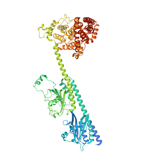
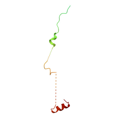
    Cone cGMP-phosphodiesterase (PDE6) is the key effector enzyme for daylight vision, and its properties are critical for shaping distinct physiology of cone photoreceptors. We determined the structures of human cone PDE6C in various liganded states by single-particle cryo-EM that reveal essential functional dynamics and adaptations of the enzyme. Our analysis exposed the dynamic nature of PDE6C association with its regulatory γ-subunit (Pγ) which allows openings of the catalytic pocket in the absence of phototransduction signaling, thereby controlling photoreceptor noise and sensitivity. We demonstrate evolutionarily recent adaptations of PDE6C stemming from residue substitutions in the Pγ subunit and the noncatalytic cGMP binding site and influencing the Pγ dynamics in holoPDE6C. Thus, our structural analysis sheds light on the previously unrecognized molecular evolution of the effector enzyme in cones that advances adaptation for photopic vision.


  • Organizational Affiliation

    Department of Molecular Physiology and Biophysics, University of Iowa Carver College of Medicine, Iowa City, IA 52242.


Macromolecules
Find similar proteins by:  (by identity cutoff)  |  3D Structure
Entity ID: 1
MoleculeChains Sequence LengthOrganismDetailsImage
Cone cGMP-specific 3',5'-cyclic phosphodiesterase subunit alpha'
A, B
843Homo sapiensMutation(s): 0 
Gene Names: PDE6CPDEA2
EC: 3.1.4.35
UniProt & NIH Common Fund Data Resources
Find proteins for P51160 (Homo sapiens)
Explore P51160 
Go to UniProtKB:  P51160
PHAROS:  P51160
GTEx:  ENSG00000095464 
Entity Groups  
Sequence Clusters30% Identity50% Identity70% Identity90% Identity95% Identity100% Identity
UniProt GroupP51160
Sequence Annotations
Expand
  • Reference Sequence
Find similar proteins by:  (by identity cutoff)  |  3D Structure
Entity ID: 2
MoleculeChains Sequence LengthOrganismDetailsImage
rod pg
C, D
99Mus musculusMutation(s): 0 
EC: 3.1.4.35
UniProt
Find proteins for P04972 (Bos taurus)
Explore P04972 
Go to UniProtKB:  P04972
Entity Groups  
Sequence Clusters30% Identity50% Identity70% Identity90% Identity95% Identity100% Identity
UniProt GroupP04972
Sequence Annotations
Expand
  • Reference Sequence
Small Molecules
Experimental Data & Validation

Experimental Data

  • Method: ELECTRON MICROSCOPY
  • Resolution: 3.00 Å
  • Aggregation State: PARTICLE 
  • Reconstruction Method: SINGLE PARTICLE 

Structure Validation

View Full Validation Report


Entry History & Funding Information

Deposition Data


Funding OrganizationLocationGrant Number
National Institutes of Health/National Eye Institute (NIH/NEI)United StatesR01 EY010843

Revision History  (Full details and data files)

  • Version 1.0: 2024-12-18
    Type: Initial release
  • Version 1.1: 2025-01-15
    Changes: Data collection, Database references