9CXE | pdb_00009cxe

SARS CoV-2 Spike protein Ectodomain with internal tag, all RBD-down conformation -C1


Experimental Data Snapshot

  • Method: ELECTRON MICROSCOPY
  • Resolution: 2.48 Å
  • Aggregation State: PARTICLE 
  • Reconstruction Method: SINGLE PARTICLE 

Starting Model: experimental
View more details

wwPDB Validation   3D Report Full Report


This is version 1.1 of the entry. See complete history


Literature

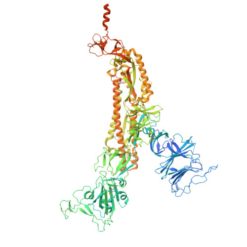
Production and cryo-electron microscopy structure of an internally tagged SARS-CoV-2 spike ecto-domain construct.

Singh, S.Liu, Y.Burke, M.Rayaprolu, V.Stein, S.E.Hasan, S.S.

(2025) J Struct Biol X 11: 100123-100123

  • DOI: https://doi.org/10.1016/j.yjsbx.2025.100123
  • Primary Citation of Related Structures:  
    9B0Y, 9B2V, 9CSS, 9CT2, 9CVH, 9CXE, 9N2L

  • PubMed Abstract: 

    The SARS-CoV-2 spike protein is synthesized in the endoplasmic reticulum of host cells, from where it undergoes export to the Golgi and the plasma membrane or retrieval from the Golgi to the endoplasmic reticulum. Elucidating the fundamental principles of this bidirectional secretion are pivotal to understanding virus assembly and designing the next generation of spike genetic vaccine with enhanced export properties. However, the widely used strategy of C-terminal affinity tagging of the spike cytosolic tail interferes with proper bidirectional trafficking. Hence, the structural and biophysical investigations of spike protein trafficking have been hindered by a lack of appropriate spike constructs. Here we describe a strategy for the internal tagging of the spike protein. Using sequence analyses and AlphaFold modeling, we identified a site down-stream of the signal sequence for the insertion of a twin-strep-tag, which facilitates purification of an ecto-domain construct from the extra-cellular medium of mammalian Expi293F cells. Mass spectrometry analyses show that the internal tag has minimal impact on N -glycan modifications, which are pivotal for spike-host interactions. Single particle cryo-electron microscopy reconstructions of the spike ecto-domain reveal conformational states compatible for ACE2 receptor interactions, further solidifying the feasibility of the internal tagging strategy. Collectively, these results present a substantial advance towards reagent development for the investigations of spike protein trafficking during coronavirus infection and genetic vaccination.


  • Organizational Affiliation

    Department of Biochemistry and Molecular Biology, University of Maryland School of Medicine, Baltimore MD 21201, USA.


Macromolecules
Find similar proteins by:  (by identity cutoff)  |  3D Structure
Entity ID: 1
MoleculeChains Sequence LengthOrganismDetailsImage
Spike glycoprotein
A, B, C
1,327Severe acute respiratory syndrome coronavirus 2Mutation(s): 6 
Gene Names: S2
UniProt
Find proteins for P0DTC2 (Severe acute respiratory syndrome coronavirus 2)
Explore P0DTC2 
Go to UniProtKB:  P0DTC2
Entity Groups  
Sequence Clusters30% Identity50% Identity70% Identity90% Identity95% Identity100% Identity
UniProt GroupP0DTC2
Glycosylation
Glycosylation Sites: 7Go to GlyGen: P0DTC2-1
Sequence Annotations
Expand
  • Reference Sequence
Oligosaccharides

Help

Entity ID: 2
MoleculeChains Length2D Diagram Glycosylation3D Interactions
2-acetamido-2-deoxy-beta-D-glucopyranose-(1-4)-[alpha-L-fucopyranose-(1-6)]2-acetamido-2-deoxy-beta-D-glucopyranoseD,
K [auth J],
R [auth P]
3N-Glycosylation
Glycosylation Resources
GlyTouCan:  G21290RB
GlyCosmos:  G21290RB
GlyGen:  G21290RB
Entity ID: 3
MoleculeChains Length2D Diagram Glycosylation3D Interactions
2-acetamido-2-deoxy-beta-D-glucopyranose-(1-4)-2-acetamido-2-deoxy-beta-D-glucopyranose
AA [auth Y],
E [auth a],
M [auth K],
N [auth L],
O [auth M],
2N-Glycosylation
Glycosylation Resources
GlyTouCan:  G42666HT
GlyCosmos:  G42666HT
GlyGen:  G42666HT
Entity ID: 4
MoleculeChains Length2D Diagram Glycosylation3D Interactions
beta-D-mannopyranose-(1-4)-2-acetamido-2-deoxy-beta-D-glucopyranose-(1-4)-2-acetamido-2-deoxy-beta-D-glucopyranose
F [auth E],
G [auth F],
I [auth H],
J [auth I],
L [auth b],
3N-Glycosylation
Glycosylation Resources
GlyTouCan:  G15407YE
GlyCosmos:  G15407YE
GlyGen:  G15407YE
Entity ID: 5
MoleculeChains Length2D Diagram Glycosylation3D Interactions
alpha-D-mannopyranose-(1-4)-2-acetamido-2-deoxy-beta-D-glucopyranose-(1-4)-2-acetamido-2-deoxy-beta-D-glucopyranoseH [auth G]3N-Glycosylation
Glycosylation Resources
GlyTouCan:  G62182OO
GlyCosmos:  G62182OO
GlyGen:  G62182OO
Small Molecules
Ligands 1 Unique
IDChains Name / Formula / InChI Key2D Diagram3D Interactions
NAG
Query on NAG

Download Ideal Coordinates CCD File 
BA [auth A]
CA [auth A]
DA [auth A]
EA [auth A]
FA [auth A]
2-acetamido-2-deoxy-beta-D-glucopyranose
C8 H15 N O6
OVRNDRQMDRJTHS-FMDGEEDCSA-N
Experimental Data & Validation

Experimental Data

  • Method: ELECTRON MICROSCOPY
  • Resolution: 2.48 Å
  • Aggregation State: PARTICLE 
  • Reconstruction Method: SINGLE PARTICLE 
EM Software:
TaskSoftware PackageVersion
MODEL REFINEMENTPHENIX1.20.1.4487-000
MODEL REFINEMENTCoot

Structure Validation

View Full Validation Report


Entry History & Funding Information

Deposition Data


Funding OrganizationLocationGrant Number
National Institutes of Health/National Institute of General Medical Sciences (NIH/NIGMS)United StatesGM150187

Revision History  (Full details and data files)

  • Version 1.0: 2025-03-05
    Type: Initial release
  • Version 1.1: 2025-03-19
    Changes: Data collection, Database references