Biophysical and Solution Structure Analysis of Critical Residues Involved in the Interaction between the PupB N-Terminal Signaling Domain and PupR C-Terminal Cell Surface Signaling Domain from Pseudomonas capeferrum.
Sultana, T., Morgan, D.M., Jernberg, B.D., Zak, P., Sinha, S.C., Colbert, C.L.(2024) Biomolecules 14
- PubMed: 39334875 Search on PubMedSearch on PubMed Central
- DOI: https://doi.org/10.3390/biom14091108
- Primary Citation Related Structures:
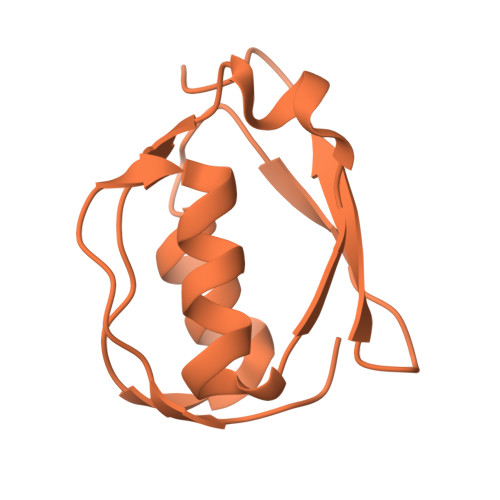
9CUV - PubMed Abstract:
Cell surface signaling (CSS) is a means of rapidly adjusting transcription in response to extracellular stimuli in Gram-negative bacteria. The pseudobactin BN7/8 uptake (Pup) system not only imports iron but also upregulates its own transcription through CSS in Pseudomonas capeferrum . In the absence of ferric pseudobactin BN7/8, the signaling components are maintained in a resting state via the formation of a periplasmic complex between the N-terminal signaling domain (NTSD) of the outer membrane iron-transporter, PupB, and the C-terminal CSS domain (CCSSD) of the sigma regulator, PupR. The previously determined 1.6 Å crystal structure of this periplasmic complex has allowed us to probe the structural and thermodynamic consequences of mutating key interfacial residues. In this report, we describe the solution structure of the PupB NTSD and use Nuclear Magnetic Resonance spectroscopy, Isothermal Titration Calorimetry, and Circular Dichroism spectroscopy together with thermal denaturation to investigate whether three PupB point mutations, Q69K, H72D, and L74A, influence the interaction merely due to the chemical nature of the amino acid substitution or also cause changes in overall protein structure. Our results demonstrate that binding to the PupR CCSSD does not alter the structure of PupB NTSD and that the individual mutations have only minor effects on structure. The mutations generally lower thermodynamic stability of the NTSD and weaken binding to the CCSSD. These findings validate the X-ray crystal structure interface, emphasizing the importance of amino acid chemical nature at the interface.
- Department of Chemistry and Biochemistry, North Dakota State University, Fargo, ND 58108, USA.
Organizational Affiliation: