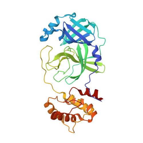
9CMS

Room-temperature X-ray structure of SARS-CoV-2 main protease drug resistant mutant (E166V) in complex with ensitrelvir (ESV)


Experimental Data Snapshot

  • Method: X-RAY DIFFRACTION
  • Resolution: 2.00 Å
  • R-Value Free: 0.195 
  • R-Value Work: 0.156 
  • R-Value Observed: 0.159 

Starting Model: experimental
View more details

wwPDB Validation   3D Report Full Report


Ligand Structure Quality Assessment 


This is version 1.0 of the entry. See complete history


Literature

Effects of SARS-CoV-2 Main Protease Mutations at Positions L50, E166, and L167 Rendering Resistance to Covalent and Noncovalent Inhibitors.

Kovalevsky, A.Aniana, A.Ghirlando, R.Coates, L.Drago, V.N.Wear, L.Gerlits, O.Nashed, N.T.Louis, J.M.

(2024) J Med Chem 

  • DOI: https://doi.org/10.1021/acs.jmedchem.4c01781
  • Primary Citation of Related Structures:  
    9CMJ, 9CMN, 9CMS, 9CMU

  • PubMed Abstract: 

    SARS-CoV-2 propagation under nirmatrelvir and ensitrelvir pressure selects for main protease (MPro) drug-resistant mutations E166V (DRM2), L50F/E166V (DRM3), E166A/L167F (DRM4), and L50F/E166A/L167F (DRM5). DRM2-DRM5 undergoes N-terminal autoprocessing to produce mature MPro with dimer dissociation constants ( K dimer ) 2-3 times larger than that of the wildtype. Co-selection of L50F restores catalytic activity of DRM2 and DRM4 from ∼10 to 30%, relative to that of the wild-type enzyme, without altering K dimer . Binding affinities and thermodynamic profiles that parallel the drug selection pressure, exhibiting significant decreases in affinity through entropy/enthalpy compensation, were compared with GC373. Reorganization of the active sites due to mutations observed in the inhibitor-free DRM3 and DRM4 structures as compared to MPro WT may account for the reduced binding affinities, although DRM2 and DRM3 complexes with ensitrelvir are almost identical to MPro WT -ensitrelvir. Chemical reactivity changes of the mutant active sites due to differences in electrostatic and protein dynamics effects likely contribute to losses in binding affinities.


  • Organizational Affiliation

    Neutron Scattering Division, Oak Ridge National Laboratory, 1 Bethel Valley Road, Oak Ridge, Tennessee 37831, United States.


Macromolecules
Find similar proteins by:  (by identity cutoff)  |  3D Structure
Entity ID: 1
MoleculeChains Sequence LengthOrganismDetailsImage
3C-like proteinase nsp5
A, B
306Severe acute respiratory syndrome coronavirus 2Mutation(s): 1 
Gene Names: rep1a-1b
EC: 3.4.22.69
UniProt
Find proteins for P0DTD1 (Severe acute respiratory syndrome coronavirus 2)
Explore P0DTD1 
Go to UniProtKB:  P0DTD1
Entity Groups  
Sequence Clusters30% Identity50% Identity70% Identity90% Identity95% Identity100% Identity
UniProt GroupP0DTD1
Sequence Annotations
Expand
  • Reference Sequence
Small Molecules
Ligands 1 Unique
IDChains Name / Formula / InChI Key2D Diagram3D Interactions
7YY (Subject of Investigation/LOI)
Query on 7YY

Download Ideal Coordinates CCD File 
C [auth A],
D [auth B]
6-[(6-chloranyl-2-methyl-indazol-5-yl)amino]-3-[(1-methyl-1,2,4-triazol-3-yl)methyl]-1-[[2,4,5-tris(fluoranyl)phenyl]methyl]-1,3,5-triazine-2,4-dione
C22 H17 Cl F3 N9 O2
QMPBBNUOBOFBFS-UHFFFAOYSA-N
Experimental Data & Validation

Experimental Data

  • Method: X-RAY DIFFRACTION
  • Resolution: 2.00 Å
  • R-Value Free: 0.195 
  • R-Value Work: 0.156 
  • R-Value Observed: 0.159 
  • Space Group: P 1 21 1
Unit Cell:
Length ( Å )Angle ( ˚ )
a = 56.195α = 90
b = 101.26β = 107.74
c = 59.374γ = 90
Software Package:
Software NamePurpose
PHENIXrefinement
CrysalisProdata reduction
CrysalisProdata scaling
PHASERphasing

Structure Validation

View Full Validation Report


Ligand Structure Quality Assessment 


Entry History & Funding Information

Deposition Data


Funding OrganizationLocationGrant Number
Not funded--

Revision History  (Full details and data files)

  • Version 1.0: 2024-10-16
    Type: Initial release