9CIN | pdb_00009cin

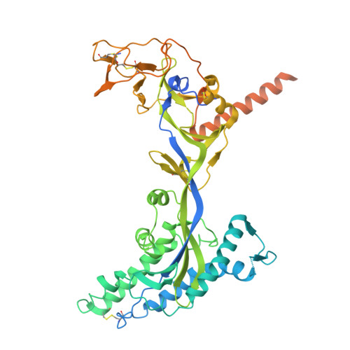
Fab fragment of Antibody with NiV glycoprotein F


Experimental Data Snapshot

  • Method: ELECTRON MICROSCOPY
  • Resolution: 3.03 Å
  • Aggregation State: PARTICLE 
  • Reconstruction Method: SINGLE PARTICLE 

wwPDB Validation 3D Report Full Report

Validation slider image for 9CIN

This is version 1.1 of the entry. See complete history

Literature

A cocktail of human mAbs targeting the henipavirus fusion and receptor binding proteins provides cross-species neutralization.

Guzman-Solis, A.A.Ouizougun-Oubari, M.Escaffre, O.Larsen, B.B.Lopez, M.Locklear, S.Kumar, M.Juelich, T.L.Smith, J.K.Zhang, L.Haas, G.D.Roenicke, R.Brambilla, L.Oguntuyo, K.Y.Patel, A.R.Sapse, I.A.Bowden, T.A.Tortorella, D.Bloom, J.D.Freiberg, A.N.Bajic, G.Duty, J.A.Lee, B.

(2026) Sci Transl Med 18: eadw8573-eadw8573

  • DOI: https://doi.org/10.1126/scitranslmed.adw8573
  • Primary Citation Related Structures: 
    9CIN

  • PubMed Abstract: 

    The Nipah and Hendra viruses (NiV and HeV, respectively) are highly pathogenic, with case fatality rates of 40 to 75%, representing substantial public health threats. Although one monoclonal antibody (mAb), mAb102.4, has advanced through phase 1 clinical trials, there remains a critical need for approved therapeutic options against these henipaviruses (HNVs). Development of human mAbs has been constrained by limited access to convalescent patient samples. Here, we describe human mAbs derived from transgenic humanized mice that cross-neutralize extant NiV and HeV strains by binding to their fusion protein (F) or receptor binding protein (RBP). Deep mutational scanning and functional studies demonstrated that the anti-RBP mAb (8G3) targets the receptor binding site and requires multiple simultaneous mutations for escape. Sequence analysis of our anti-F mAbs identified a clonally expanded VH3-33 family with evidence of somatic hypermutation, yielding high-affinity antibodies. Cryo-electron microscopy revealed that our most potent F antibody (2A1) recognizes a conserved quaternary epitope spanning two protomers in trimeric prefusion NiV-F and stabilized, rather than displaced, a key glycan shield, distinguishing it from previously described antibodies targeting this region. The 8G3 and 2A1 mAbs exhibited additive neutralization when combined and provided complete protection against lethal NiV challenge in hamsters when administered individually or as a cocktail, even when treatment was delayed. Using a pseudovirus system, we show that this dual-targeting approach was resilient against a suite of escape mutants compared with monotherapy. Our findings establish a candidate therapeutic strategy that minimizes development of resistance, providing a foundation for next-generation countermeasures against emerging HNVs.


  • Organizational Affiliation
    • Department of Microbiology, Icahn School of Medicine at Mount Sinai, New York, NY 10029, USA.

Macromolecule Content 

  • Total Structure Weight: 261.94 kDa 
  • Atom Count: 15,798 
  • Modeled Residue Count: 2,007 
  • Deposited Residue Count: 2,343 
  • Unique protein chains: 3

Macromolecules

Find similar proteins by:|  3D Structure
Entity ID: 1
MoleculeChains  Sequence LengthOrganismDetailsImage
Fusion glycoprotein F0A,
F [auth B],
G [auth C]
552Henipavirus nipahenseMutation(s): 0 
UniProt
Find proteins for Q9IH63 (Nipah virus)
Explore Q9IH63 
Go to UniProtKB:  Q9IH63
Entity Groups
Sequence Clusters30% Identity50% Identity70% Identity90% Identity95% Identity100% Identity
UniProt GroupQ9IH63
Glycosylation
Glycosylation Sites: 4
Sequence Annotations
Expand
Reference Sequence
Find similar proteins by:|  3D Structure
Entity ID: 2
MoleculeChains  Sequence LengthOrganismDetailsImage
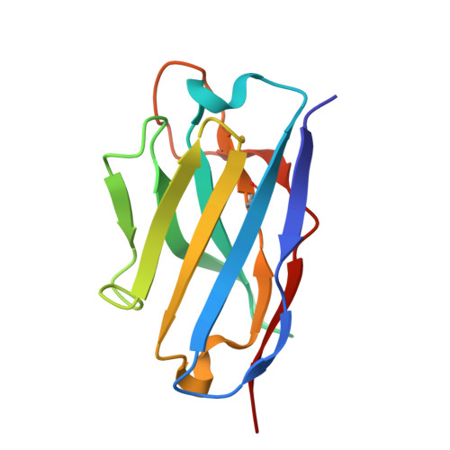
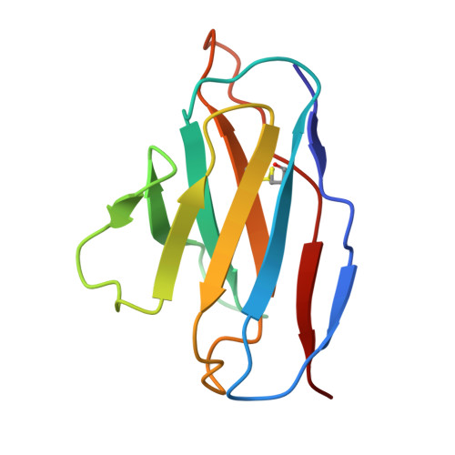
Antibody Heavy ChainB [auth H],
C [auth J],
D [auth M]
122Mus musculusMutation(s): 0 
Entity Groups
Sequence Clusters30% Identity50% Identity70% Identity90% Identity95% Identity100% Identity
Sequence Annotations
Expand
Reference Sequence
Find similar proteins by:|  3D Structure
Entity ID: 3
MoleculeChains  Sequence LengthOrganismDetailsImage
Antibody Light ChainE [auth L],
H [auth K],
I [auth N]
107Mus musculusMutation(s): 0 
Entity Groups
Sequence Clusters30% Identity50% Identity70% Identity90% Identity95% Identity100% Identity
Sequence Annotations
Expand
Reference Sequence

Oligosaccharides

Help  
Entity ID: 4
MoleculeChains Length2D Diagram GlycosylationD Interactions
2-acetamido-2-deoxy-beta-D-glucopyranose-(1-4)-2-acetamido-2-deoxy-beta-D-glucopyranoseJ [auth D],
L [auth F],
N [auth I]
2N-Glycosylation
Glycosylation Resources
GlyTouCan: G42666HT
GlyCosmos: G42666HT
GlyGen: G42666HT
Entity ID: 5
MoleculeChains Length2D Diagram GlycosylationD Interactions
alpha-D-mannopyranose-(1-3)-beta-D-mannopyranose-(1-4)-2-acetamido-2-deoxy-beta-D-glucopyranose-(1-4)-2-acetamido-2-deoxy-beta-D-glucopyranoseK [auth E],
M [auth G],
O
4N-Glycosylation
Glycosylation Resources
GlyTouCan: G81315DD
GlyCosmos: G81315DD
GlyGen: G81315DD

Experimental Data & Validation

Experimental Data

  • Method: ELECTRON MICROSCOPY
  • Resolution: 3.03 Å
  • Aggregation State: PARTICLE 
  • Reconstruction Method: SINGLE PARTICLE 
EM Software:
TaskSoftware PackageVersion
MODEL REFINEMENTPHENIX1.21.2_5419+SVN
RECONSTRUCTIONcryoSPARC

Structure Validation

View Full Validation Report


Entry History 

& Funding Information

Deposition Data


Funding OrganizationLocationGrant Number
Not funded--

Revision History  (Full details and data files)

  • Version 1.0: 2026-01-28
    Type: Initial release
  • Version 1.1: 2026-08-12
    Changes: Data collection, Database references