9CHL | pdb_00009chl

P. vulgaris tetrameric HigBA- operator 2 DNA


Experimental Data Snapshot

  • Method: X-RAY DIFFRACTION
  • Resolution: 2.40 Å
  • R-Value Free: 
    0.221 (Depositor), 0.224 (DCC) 
  • R-Value Work: 
    0.175 (Depositor), 0.178 (DCC) 

wwPDB Validation   3D Report Full Report


This is version 1.1 of the entry. See complete history


Literature

Antitoxin control of optimal transcriptional repression in the atypical HigB-HigA toxin-antitoxin system from Proteus vulgaris.

Pavelich, I.J.Schureck, M.A.Srinivas, P.Blackburn, T.M.Wang, D.Hoffer, E.D.Boamah, M.Zaldana, K.Onuoha, N.Miles, S.J.Grabowicz, M.Okafor, C.D.Dunham, C.M.

(2025) Nucleic Acids Res 53

  • DOI: https://doi.org/10.1093/nar/gkaf610
  • Primary Citation of Related Structures:  
    9CHL, 9CHN

  • PubMed Abstract: 

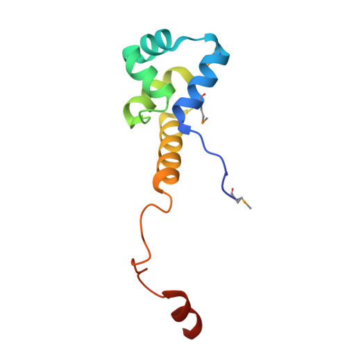
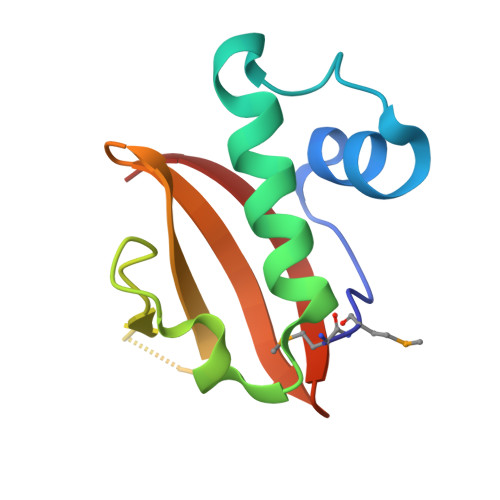
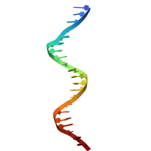
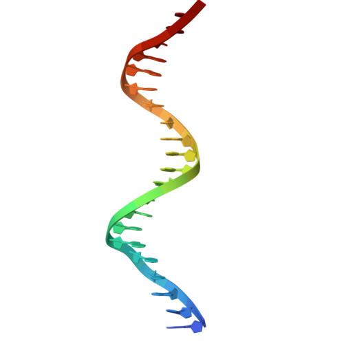
    Bacterial toxin-antitoxin (TA) pairs transcriptionally autoregulate their expression via a repression/derepression mechanism in response to changing environmental conditions. The structural diversity of TA systems influences the mechanisms of transcriptional regulation. Here, we define the molecular mechanism for the plasmid-encoded HigB-HigA TA pair originally identified in a post-operative infection with antibiotic-resistant Proteus vulgaris. We determine DNA binding and promoter activity by the HigB-HigA complex supported by structural biology and molecular dynamics simulations of an elusive DNA operator-TA repressor complex. To define the optimal oligomeric TA repressor-DNA operator complex required for derepression, we engineered a dedicated trimeric HigB-HigA2 complex that represses transcription more than 26-fold as compared to the tetrameric HigB2-HigA2. These results expand the known diversity of how the HigB-HigA TA family is autoregulated.


  • Organizational Affiliation
    • Department of Chemistry and the Emory Antibiotic Resistance Center, Emory University, Atlanta, GA 30322, United States.

Macromolecules

Find similar proteins by:  (by identity cutoff)  |  3D Structure
Entity ID: 1
MoleculeChains Sequence LengthOrganismDetailsImage
Antitoxin HigAA,
C,
G [auth E],
I [auth G]
104Proteus vulgarisMutation(s): 0 
Gene Names: higA
UniProt
Find proteins for Q7A224 (Proteus vulgaris)
Explore Q7A224 
Go to UniProtKB:  Q7A224
Entity Groups  
Sequence Clusters30% Identity50% Identity70% Identity90% Identity95% Identity100% Identity
UniProt GroupQ7A224
Sequence Annotations
Expand
  • Reference Sequence
Find similar proteins by:  (by identity cutoff)  |  3D Structure
Entity ID: 2
MoleculeChains Sequence LengthOrganismDetailsImage
Endoribonuclease HigBB,
D,
H [auth F],
J [auth H]
93Proteus vulgarisMutation(s): 0 
Gene Names: higB
EC: 3.1
UniProt
Find proteins for Q7A225 (Proteus vulgaris)
Explore Q7A225 
Go to UniProtKB:  Q7A225
Entity Groups  
Sequence Clusters30% Identity50% Identity70% Identity90% Identity95% Identity100% Identity
UniProt GroupQ7A225
Sequence Annotations
Expand
  • Reference Sequence

Find similar nucleic acids by:  Sequence   |   3D Structure  

Entity ID: 3
MoleculeChains LengthOrganismImage
DNA (5'-D(*GP*TP*AP*TP*TP*AP*CP*AP*CP*AP*CP*CP*AP*TP*GP*TP*AP*AP*TP*AP*C)-3')E [auth I],
K
21Proteus vulgaris
Sequence Annotations
Expand
  • Reference Sequence

Find similar nucleic acids by:  Sequence   |   3D Structure  

Entity ID: 4
MoleculeChains LengthOrganismImage
DNA (5'-D(*GP*TP*AP*TP*TP*AP*CP*AP*TP*GP*GP*TP*GP*TP*GP*TP*AP*AP*TP*AP*C)-3')F [auth J],
L
21Proteus vulgaris
Sequence Annotations
Expand
  • Reference Sequence
Small Molecules
Ligands 2 Unique
IDChains Name / Formula / InChI Key2D Diagram3D Interactions
CL
Query on CL

Download Ideal Coordinates CCD File 
AA [auth H],
N [auth B],
P [auth C],
R [auth D],
W [auth F]
CHLORIDE ION
Cl
VEXZGXHMUGYJMC-UHFFFAOYSA-M
MG
Query on MG

Download Ideal Coordinates CCD File 
BA [auth K]
CA [auth K]
DA [auth K]
EA [auth L]
M [auth A]
BA [auth K],
CA [auth K],
DA [auth K],
EA [auth L],
M [auth A],
O [auth B],
Q [auth C],
S [auth J],
T [auth J],
U [auth J],
V [auth E],
X [auth G],
Y [auth G],
Z [auth H]
MAGNESIUM ION
Mg
JLVVSXFLKOJNIY-UHFFFAOYSA-N
Modified Residues  1 Unique
IDChains TypeFormula2D DiagramParent
MSE
Query on MSE
A,
C,
G [auth E],
I [auth G]
L-PEPTIDE LINKINGC5 H11 N O2 SeMET
Experimental Data & Validation

Experimental Data

  • Method: X-RAY DIFFRACTION
  • Resolution: 2.40 Å
  • R-Value Free:  0.221 (Depositor), 0.224 (DCC) 
  • R-Value Work:  0.175 (Depositor), 0.178 (DCC) 
Space Group: C 1 2 1
Unit Cell:
Length ( Å )Angle ( ˚ )
a = 98.15α = 90
b = 98.13β = 109.49
c = 147.19γ = 90
Software Package:
Software NamePurpose
XDSdata reduction
XSCALEdata scaling
PHASERphasing
PHENIXrefinement
Cootmodel building

Structure Validation

View Full Validation Report


Entry History & Funding Information

Deposition Data


Funding OrganizationLocationGrant Number
Other governmentUnited States--
Other privateUnited States--

Revision History  (Full details and data files)

  • Version 1.0: 2025-07-02
    Type: Initial release
  • Version 1.1: 2026-01-14
    Changes: Database references