9CG1 | pdb_00009cg1

DUF512 protein from Clostridium sporogenes


Experimental Data Snapshot

  • Method: X-RAY DIFFRACTION
  • Resolution: 1.68 Å
  • R-Value Free: 
    0.198 (Depositor), 0.198 (DCC) 
  • R-Value Work: 
    0.158 (Depositor), 0.158 (DCC) 
  • R-Value Observed: 
    0.160 (Depositor) 

Starting Model: in silico
View more details

wwPDB Validation 3D Report Full Report

Validation slider image for 9CG1

Ligand Structure Quality Assessment 


This is version 1.1 of the entry. See complete history

Literature

Structural Evidence for DUF512 as a Radical S -Adenosylmethionine Cobalamin-Binding Domain.

Wang, B.Solinski, A.E.Radle, M.I.Peduzzi, O.M.Knox, H.L.Cui, J.Maurya, R.K.Yennawar, N.H.Booker, S.J.

(2024) ACS Bio Med Chem Au 4: 319-330

  • DOI: https://doi.org/10.1021/acsbiomedchemau.4c00067
  • Primary Citation Related Structures: 
    9CG1, 9CG2

  • PubMed Abstract: 

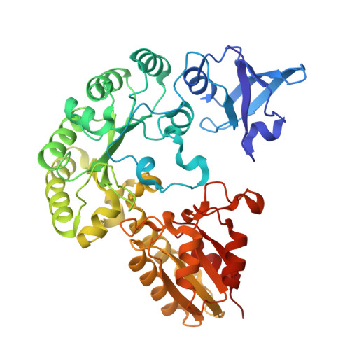
    Cobalamin (Cbl)-dependent radical S -adenosylmethionine (SAM) enzymes constitute a large subclass of radical SAM (RS) enzymes that use Cbl to catalyze various types of reactions, the most common of which are methylations. Most Cbl-dependent RS enzymes contain an N-terminal Rossmann fold that aids Cbl binding. Recently, it has been demonstrated that the methanogenesis marker protein 10 (Mmp10) requires Cbl to methylate an arginine residue in the α-subunit of methyl coenzyme M reductase. However, Mmp10 contains a Cbl-binding domain in the C-terminal region of its primary structure that does not share significant sequence similarity with canonical RS Cbl-binding domains. Bioinformatic analysis of Mmp10 identified DUF512 (Domain of Unknown Function 512) as a potential Cbl-binding domain in RS enzymes. In this paper, four randomly selected DUF512-containing proteins from various organisms were overexpressed, purified, and shown to bind Cbl. X-ray crystal structures of DUF512-containing proteins from Clostridium sporogenes and Pyrococcus furiosus were determined, confirming their C-terminal Cbl-binding domains. The structure of the DUF512-containing protein from C. sporogenes is the first of an RS enzyme containing a PDZ domain. Its RS domain has an unprecedented β 3 α 4 core, whereas most RS enzymes adopt a (βα) 6 core. The DUF512-containing protein from P. furiosus has no PDZ domain, but its RS domain also has an uncommon (βα) 5 core.


  • Organizational Affiliation
    • Department of Chemistry, The Pennsylvania State University, University Park, Pennsylvania 16802, United States.

Macromolecule Content 

  • Total Structure Weight: 53.86 kDa 
  • Atom Count: 4,359 
  • Modeled Residue Count: 443 
  • Deposited Residue Count: 443 
  • Unique protein chains: 1

Macromolecules

Find similar proteins by:|  3D Structure
Entity ID: 1
MoleculeChains  Sequence LengthOrganismDetailsImage
Radical SAM protein443Clostridium sporogenesMutation(s): 0 
Gene Names: CGS26_13040FDB81_14220

Small Molecules

Ligands 6 Unique
IDChains Name / Formula / InChI Key2D Diagram3D Interactions
B12
(Subject of Investigation/LOI)

Query on B12


Download:Ideal Coordinates CCD File
E [auth A]COBALAMIN
C62 H89 Co N13 O14 P
LKVIQTCSMMVGFU-DWSMJLPVSA-N
SA8
(Subject of Investigation/LOI)

Query on SA8


Download:Ideal Coordinates CCD File
H [auth A]S-5'-AZAMETHIONINE-5'-DEOXYADENOSINE
C15 H23 N7 O5
JISVTSUBJCPLSV-TWBCTODHSA-N
SF4
(Subject of Investigation/LOI)

Query on SF4


Download:Ideal Coordinates CCD File
F [auth A]IRON/SULFUR CLUSTER
Fe4 S4
LJBDFODJNLIPKO-UHFFFAOYSA-N
GOL

Query on GOL


Download:Ideal Coordinates CCD File
B [auth A]GLYCEROL
C3 H8 O3
PEDCQBHIVMGVHV-UHFFFAOYSA-N
IMD

Query on IMD


Download:Ideal Coordinates CCD File
G [auth A],
I [auth A]
IMIDAZOLE
C3 H5 N2
RAXXELZNTBOGNW-UHFFFAOYSA-O
BO3

Query on BO3


Download:Ideal Coordinates CCD File
C [auth A],
D [auth A]
BORIC ACID
B H3 O3
KGBXLFKZBHKPEV-UHFFFAOYSA-N

Experimental Data & Validation

Experimental Data

  • Method: X-RAY DIFFRACTION
  • Resolution: 1.68 Å
  • R-Value Free:  0.198 (Depositor), 0.198 (DCC) 
  • R-Value Work:  0.158 (Depositor), 0.158 (DCC) 
  • R-Value Observed: 0.160 (Depositor) 
Space Group: P 1 21 1
Unit Cell:
Length ( Å )Angle ( ˚ )
a = 56.67α = 90
b = 59.302β = 109.82
c = 81.059γ = 90
Software Package:
Software NamePurpose
PHENIXrefinement
HKL-2000data reduction
HKL-2000data scaling
PHASERphasing

Structure Validation

View Full Validation Report


Ligand Structure Quality Assessment 


Entry History 

& Funding Information

Deposition Data


Funding OrganizationLocationGrant Number
National Institutes of Health/National Institute of General Medical Sciences (NIH/NIGMS)United StatesGM-122595

Revision History  (Full details and data files)

  • Version 1.0: 2025-03-05
    Type: Initial release
  • Version 1.1: 2025-09-17
    Changes: Database references