9CEG | pdb_00009ceg

20S Proteasome core particle beta-T1A mutant resting state (Frame 20)


Experimental Data Snapshot

  • Method: ELECTRON MICROSCOPY
  • Resolution: 2.86 Å
  • Aggregation State: PARTICLE 
  • Reconstruction Method: SINGLE PARTICLE 

wwPDB Validation   3D Report Full Report


This is version 1.1 of the entry. See complete history


Literature

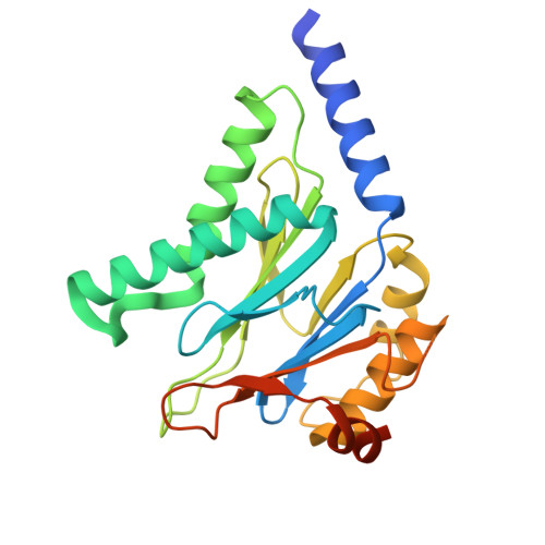
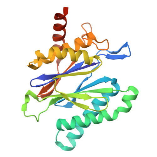
Structural basis for allosteric modulation of M. tuberculosis proteasome core particle.

Turner, M.Uday, A.B.Velyvis, A.Rennella, E.Zeytuni, N.Vahidi, S.

(2025) Nat Commun 16: 3138-3138

  • DOI: https://doi.org/10.1038/s41467-025-58430-0
  • Primary Citation of Related Structures:  
    9CE5, 9CE7, 9CE8, 9CEB, 9CEE, 9CEG

  • PubMed Abstract: 

    The Mycobacterium tuberculosis (Mtb) proteasome system selectively degrades damaged or misfolded proteins and is crucial for the pathogen's survival within the host. Targeting the 20S core particle (CP) offers a viable strategy for developing tuberculosis treatments. The activity of Mtb 20S CP, like that of its eukaryotic counterpart, is allosterically regulated, yet the specific conformations involved have not been captured in high-resolution structures to date. Here, we use single-particle electron cryomicroscopy and H/D exchange mass spectrometry to determine the Mtb 20S CP structure in an auto-inhibited state that is distinguished from the canonical resting state by the conformation of switch helices at the α/β interface. The rearrangement of these helices collapses the S1 pocket, effectively inhibiting substrate binding. Biochemical experiments show that the Mtb 20S CP activity can be altered through allosteric sites far from the active site. Our findings underscore the potential of targeting allostery to develop antituberculosis therapeutics.


  • Organizational Affiliation
    • Department of Molecular and Cellular Biology, University of Guelph, Guelph, Ontario, Canada.

Macromolecules
Find similar proteins by:  (by identity cutoff)  |  3D Structure
Entity ID: 1
MoleculeChains Sequence LengthOrganismDetailsImage
Proteasome subunit alpha
A, B, C, D, E
A, B, C, D, E, F, G, H, I, J, K, L, M, N
248Mycobacterium tuberculosisMutation(s): 0 
Gene Names: prcARv2109c
UniProt
Find proteins for P9WHU1 (Mycobacterium tuberculosis (strain ATCC 25618 / H37Rv))
Explore P9WHU1 
Go to UniProtKB:  P9WHU1
Entity Groups  
Sequence Clusters30% Identity50% Identity70% Identity90% Identity95% Identity100% Identity
UniProt GroupP9WHU1
Sequence Annotations
Expand
  • Reference Sequence
Find similar proteins by:  (by identity cutoff)  |  3D Structure
Entity ID: 2
MoleculeChains Sequence LengthOrganismDetailsImage
Proteasome subunit beta234Mycobacterium tuberculosisMutation(s): 1 
Gene Names: prcBRv2110c
EC: 3.4.25.1
UniProt
Find proteins for P9WHT9 (Mycobacterium tuberculosis (strain ATCC 25618 / H37Rv))
Explore P9WHT9 
Go to UniProtKB:  P9WHT9
Entity Groups  
Sequence Clusters30% Identity50% Identity70% Identity90% Identity95% Identity100% Identity
UniProt GroupP9WHT9
Sequence Annotations
Expand
  • Reference Sequence
Experimental Data & Validation

Experimental Data

  • Method: ELECTRON MICROSCOPY
  • Resolution: 2.86 Å
  • Aggregation State: PARTICLE 
  • Reconstruction Method: SINGLE PARTICLE 
EM Software:
TaskSoftware PackageVersion
MODEL REFINEMENTPHENIXdev_5316
RECONSTRUCTIONcryoSPARC4

Structure Validation

View Full Validation Report


Entry History & Funding Information

Deposition Data


Funding OrganizationLocationGrant Number
Canadian Institutes of Health Research (CIHR)CanadaPJT451412
Natural Sciences and Engineering Research Council (NSERC, Canada)CanadaRGPIN/03031-2022

Revision History  (Full details and data files)

  • Version 1.0: 2025-03-12
    Type: Initial release
  • Version 1.1: 2025-04-16
    Changes: Data collection, Database references