Key structural role of a conserved cis-proline revealed by the P285S variant of soybean serine hydroxymethyltransferase 8.
Samarakoon, V., Owuocha, L.F., Hammond, J., Mitchum, M.G., Beamer, L.J.(2024) Biochem J 481: 1557-1568
- PubMed: 39373197
- DOI: https://doi.org/10.1042/BCJ20240338
- Primary Citation of Related Structures:
9CE6, 9CG8 - PubMed Abstract:
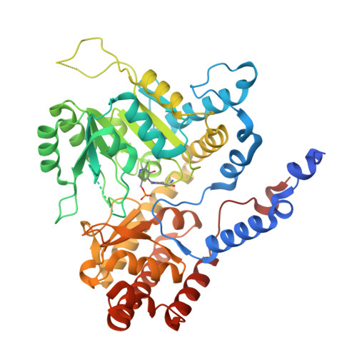
The enzyme serine hydroxymethyltransferase (SHMT) plays a key role in folate metabolism and is conserved in all kingdoms of life. SHMT is a pyridoxal 5'-phosphate (PLP) - dependent enzyme that catalyzes the conversion of L-serine and (6S)-tetrahydrofolate to glycine and 5,10-methylene tetrahydrofolate. Crystal structures of multiple members of the SHMT family have shown that the enzyme has a single conserved cis proline, which is located near the active site. Here, we have characterized a Pro to Ser amino acid variant (P285S) that affects this conserved cis proline in soybean SHMT8. P285S was identified as one of a set of mutations that affect the resistance of soybean to the agricultural pathogen soybean cyst nematode. We find that replacement of Pro285 by serine eliminates PLP-mediated catalytic activity of SHMT8, reduces folate binding, decreases enzyme stability, and affects the dimer-tetramer ratio of the enzyme in solution. Crystal structures at 1.9 - 2.2 Å resolution reveal a local reordering of the polypeptide chain that extends an a-helix and shifts a turn region into the active site. This results in a dramatically perturbed PLP-binding pose, where the ring of the cofactor is flipped by ~180° with concomitant loss of conserved enzyme-PLP interactions. A nearby region of the polypeptide becomes disordered, evidenced by missing electron density for ~10 residues. These structural perturbations are consistent with the loss of enzyme activity and folate binding and underscore the important role of the Pro285 cis-peptide in SHMT structure and function.
- university of missouri, Columbia, Missouri, United States.
Organizational Affiliation:


