9CE0 | pdb_00009ce0

DosP Apo Bent form


Experimental Data Snapshot

  • Method: ELECTRON MICROSCOPY
  • Resolution: 3.97 Å
  • Aggregation State: PARTICLE 
  • Reconstruction Method: SINGLE PARTICLE 

wwPDB Validation 3D Report Full Report

Validation slider image for 9CE0

This is version 1.2 of the entry. See complete history

Literature

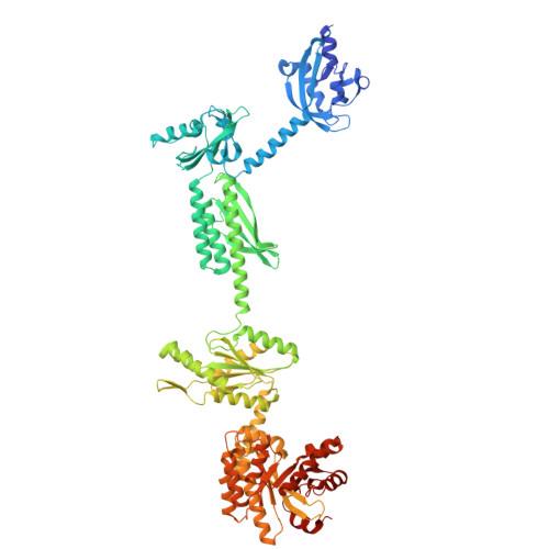
Structures of the multi-domain oxygen sensor DosP: remote control of a c-di-GMP phosphodiesterase by a regulatory PAS domain.

Wu, W.Kumar, P.Brautigam, C.A.Tso, S.C.Baniasadi, H.R.Kober, D.L.Gilles-Gonzalez, M.A.

(2024) Nat Commun 15: 9653-9653

  • DOI: https://doi.org/10.1038/s41467-024-53942-7
  • Primary Citation Related Structures: 
    9BGV, 9BKV, 9CDR, 9CE0, 9CLO, 9CMF

  • PubMed Abstract: 

    The heme-based direct oxygen sensor DosP degrades c-di-GMP, a second messenger nearly unique to bacteria. In stationary phase Escherichia coli, DosP is the most abundant c-di-GMP phosphodiesterase. Ligation of O 2 to a heme-binding PAS domain (hPAS) of the protein enhances the phosphodiesterase through an allosteric mechanism that has remained elusive. We determine six structures of full-length DosP in its aerobic or anaerobic conformations, with or without c-di-GMP. DosP is an elongated dimer with the regulatory heme containing domain and phosphodiesterase separated by nearly 180 Å. In the absence of substrate, regardless of the heme status, DosP presents an equilibrium of two distinct conformations. Binding of substrate induces DosP to adopt a single, ON-state or OFF-state conformation depending on its heme status. Structural and biochemical studies of this multi-domain sensor and its mutants provide insights into signal regulation of second-messenger levels.


  • Organizational Affiliation
    • Department of Biochemistry, University of Texas Southwestern Medical Center, Dallas, TX, 75390, USA.

Macromolecule Content 

  • Total Structure Weight: 183.61 kDa 
  • Atom Count: 12,551 
  • Modeled Residue Count: 1,568 
  • Deposited Residue Count: 1,612 
  • Unique protein chains: 1

Macromolecules

Find similar proteins by:|  3D Structure
Entity ID: 1
MoleculeChains  Sequence LengthOrganismDetailsImage
Oxygen sensor protein DosP
A, B
806Escherichia coliMutation(s): 0 
Gene Names: dosPdospdeOyddUb1489JW1484
EC: 3.1.4.52
UniProt
Find proteins for P76129 (Escherichia coli (strain K12))
Explore P76129 
Go to UniProtKB:  P76129
Entity Groups
Sequence Clusters30% Identity50% Identity70% Identity90% Identity95% Identity100% Identity
UniProt GroupP76129
Sequence Annotations
Expand
Reference Sequence

Experimental Data & Validation

Experimental Data

  • Method: ELECTRON MICROSCOPY
  • Resolution: 3.97 Å
  • Aggregation State: PARTICLE 
  • Reconstruction Method: SINGLE PARTICLE 
EM Software:
TaskSoftware PackageVersion
MODEL REFINEMENTPHENIX

Structure Validation

View Full Validation Report


Entry History 

& Funding Information

Deposition Data


Funding OrganizationLocationGrant Number
National Institutes of Health/National Institute of General Medical Sciences (NIH/NIGMS)United StatesR00 GM141261
National Institutes of Health/National Institute of General Medical Sciences (NIH/NIGMS)United StatesU24 GM129547

Revision History  (Full details and data files)

  • Version 1.0: 2024-11-20
    Type: Initial release
  • Version 1.1: 2024-11-27
    Changes: Data collection, Database references
  • Version 1.2: 2025-05-14
    Changes: Data collection