9CDQ | pdb_00009cdq

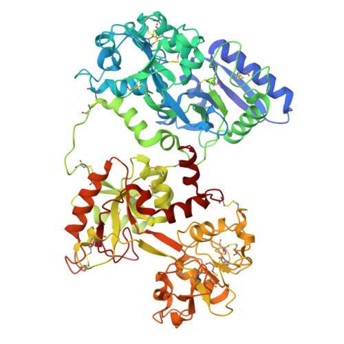
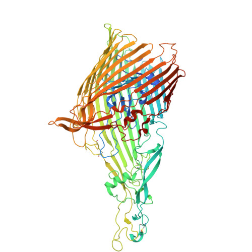
Transferrin Binding Protein A in complex with transferrin (iron bound in N lobe only)


Experimental Data Snapshot

  • Method: ELECTRON MICROSCOPY
  • Resolution: 3.76 Å
  • Aggregation State: PARTICLE 
  • Reconstruction Method: SINGLE PARTICLE 

wwPDB Validation 3D Report Full Report

Validation slider image for 9CDQ

This is version 1.1 of the entry. See complete history

Literature

Structural insights into the mechanism underpinning iron piracy in pathogenic Neisseria.

Dubey, S.Stoudenmire, J.Bury, G.Yang, L.Fan, Z.Li, P.Wadhwa, G.Pushkar, Y.Gumbart, J.C.Cornelissen, C.N.Noinaj, N.

(2026) Sci Adv 12: eaea2470-eaea2470

  • DOI: https://doi.org/10.1126/sciadv.aea2470
  • Primary Citation Related Structures: 
    9CDQ, 9CLT

  • PubMed Abstract: 

    The pathogenesis of Neisseria hinges on the surface proteins TbpA and TbpB, which orchestrate the acquisition of iron from transferrin. TbpB selectively captures iron-loaded transferrin and delivers it to TbpA for iron import. We report a series of cryo-electron microscopy structures of trapped intermediates along the iron acquisition pathway. These structural studies are supported by pulldowns, electron paramagnetic resonance studies, molecular dynamics simulations, and studies in Neisseria gonorrhoeae , which show that TbpA mechanically opens the C-lobe of transferrin, triggering iron release. Once iron is removed, TbpB dissociates and undergoes large subunit rearrangements with its C-lobe rebinding at a different interface on transferrin. TonB binding expands the barrel of TbpA, helping displace the plug to open a path for iron import. This also disrupts the interaction of the plug loop with the C1 domain of transferrin, leading to the dissociation of the spent transferrin. Together, our study provides a more complete understanding of metal acquisition systems in Neisseria and other Gram-negative bacteria.


  • Organizational Affiliation
    • Markey Center for Structural Biology, Department of Biological Sciences, Purdue University, West Lafayette, IN 47907, USA.

Macromolecule Content 

  • Total Structure Weight: 179.73 kDa 
  • Atom Count: 12,048 
  • Modeled Residue Count: 1,545 
  • Deposited Residue Count: 1,613 
  • Unique protein chains: 2

Macromolecules

Find similar proteins by:|  3D Structure
Entity ID: 1
MoleculeChains  Sequence LengthOrganismDetailsImage
SerotransferrinA [auth F]698Homo sapiensMutation(s): 0 
UniProt & NIH Common Fund Data Resources
Find proteins for P02787 (Homo sapiens)
Explore P02787 
Go to UniProtKB:  P02787
PHAROS:  P02787
GTEx:  ENSG00000091513 
Entity Groups
Sequence Clusters30% Identity50% Identity70% Identity90% Identity95% Identity100% Identity
UniProt GroupP02787
Sequence Annotations
Expand
Reference Sequence
Find similar proteins by:|  3D Structure
Entity ID: 2
MoleculeChains  Sequence LengthOrganismDetailsImage
Transferrin-binding protein AB [auth A]915Neisseria meningitidis serogroup BMutation(s): 0 
Gene Names: tbp1tbpANMB0461
UniProt
Find proteins for Q9K0U9 (Neisseria meningitidis serogroup B (strain ATCC BAA-335 / MC58))
Explore Q9K0U9 
Go to UniProtKB:  Q9K0U9
Entity Groups
Sequence Clusters30% Identity50% Identity70% Identity90% Identity95% Identity100% Identity
UniProt GroupQ9K0U9
Sequence Annotations
Expand
Reference Sequence

Experimental Data & Validation

Experimental Data

  • Method: ELECTRON MICROSCOPY
  • Resolution: 3.76 Å
  • Aggregation State: PARTICLE 
  • Reconstruction Method: SINGLE PARTICLE 

Structure Validation

View Full Validation Report


Entry History 

& Funding Information

Deposition Data


Funding OrganizationLocationGrant Number
National Institutes of Health/National Institute Of Allergy and Infectious Diseases (NIH/NIAID)United States1R01AI127793

Revision History  (Full details and data files)

  • Version 1.0: 2026-03-11
    Type: Initial release
  • Version 1.1: 2026-04-15
    Changes: Data collection, Database references