9C91 | pdb_00009c91

Assimilatory NADPH-dependent sulfite reductase minimal dimer


Experimental Data Snapshot

  • Method: ELECTRON MICROSCOPY
  • Resolution: 2.78 Å
  • Aggregation State: PARTICLE 
  • Reconstruction Method: SINGLE PARTICLE 

wwPDB Validation 3D Report Full Report

Validation slider image for 9C91

This is version 1.3 of the entry. See complete history

Literature

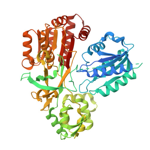
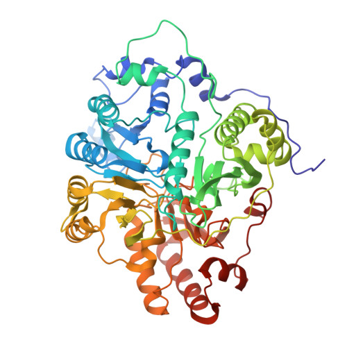
Structure of dimerized assimilatory NADPH-dependent sulfite reductase reveals the minimal interface for diflavin reductase binding.

Ghazi Esfahani, B.Walia, N.Neselu, K.Garg, Y.Aragon, M.Askenasy, I.Wei, H.A.Mendez, J.H.Stroupe, M.E.

(2025) Nat Commun 16: 2955-2955

  • DOI: https://doi.org/10.1038/s41467-025-58037-5
  • Primary Citation Related Structures: 
    9C91

  • PubMed Abstract: 

    Escherichia coli NADPH-dependent assimilatory sulfite reductase (SiR) reduces sulfite by six electrons to make sulfide for incorporation into sulfur-containing biomolecules. SiR has two subunits: an NADPH, FMN, and FAD-binding diflavin flavoprotein and a siroheme/Fe 4 S 4 cluster-containing hemoprotein. The molecular interactions that govern subunit binding have been unknown since the discovery of SiR over 50 years ago because SiR is flexible, thus has been intransigent for traditional high-resolution structural analysis. We use a combination of the chameleon® plunging system with a fluorinated lipid to overcome the challenges of preserving a flexible molecule to determine a 2.78 Å-resolution cryo-EM structure of a minimal heterodimer complex. Chameleon®, combined with the fluorinated lipid, overcomes persistent denaturation at the air-water interface. Using a previously characterized minimal heterodimer reduces the heterogeneity of a structurally heterogeneous complex to a level that we analyze using multi-conformer cryo-EM image analysis algorithms. Here, we report the near-atomic resolution structure of the flavoprotein/hemoprotein complex, revealing how they interact in a minimal interface. Further, we determine the structural elements that discriminate between pairing a hemoprotein with a diflavin reductase, as in the E. coli homolog, or a ferredoxin partner, as in maize (Zea mays).


  • Organizational Affiliation
    • Department of Biological Science and Institute of Molecular Biophysics, Florida State University, Tallahassee, FL, USA.

Macromolecule Content 

  • Total Structure Weight: 130.83 kDa 
  • Atom Count: 8,784 
  • Modeled Residue Count: 1,088 
  • Deposited Residue Count: 1,145 
  • Unique protein chains: 2

Macromolecules

Find similar proteins by:|  3D Structure
Entity ID: 1
MoleculeChains  Sequence LengthOrganismDetailsImage
Sulfite reductase [NADPH] flavoprotein alpha-component575Escherichia coliMutation(s): 0 
Gene Names: cysJHEP34_003108
EC: 1.8.1.2
UniProt
Find proteins for P38038 (Escherichia coli (strain K12))
Explore P38038 
Go to UniProtKB:  P38038
Entity Groups
Sequence Clusters30% Identity50% Identity70% Identity90% Identity95% Identity100% Identity
UniProt GroupP38038
Sequence Annotations
Expand
Reference Sequence
Find similar proteins by:|  3D Structure
Entity ID: 2
MoleculeChains  Sequence LengthOrganismDetailsImage
Sulfite reductase [NADPH] hemoprotein beta-component570Escherichia coliMutation(s): 0 
Gene Names: cysIBANRA_01280HMV95_03860IH772_07600P6223_002112
EC: 1.8.1.2
UniProt
Find proteins for P17846 (Escherichia coli (strain K12))
Explore P17846 
Go to UniProtKB:  P17846
Entity Groups
Sequence Clusters30% Identity50% Identity70% Identity90% Identity95% Identity100% Identity
UniProt GroupP17846
Sequence Annotations
Expand
Reference Sequence

Small Molecules

Ligands 6 Unique
IDChains Name / Formula / InChI Key2D Diagram3D Interactions
SRM

Query on SRM


Download:Ideal Coordinates CCD File
H [auth B]SIROHEME
C42 H44 Fe N4 O16
DLKSSIHHLYNIKN-MWBYXLBFSA-L
FAD

Query on FAD


Download:Ideal Coordinates CCD File
C [auth A]FLAVIN-ADENINE DINUCLEOTIDE
C27 H33 N9 O15 P2
VWWQXMAJTJZDQX-UYBVJOGSSA-N
FMN

Query on FMN


Download:Ideal Coordinates CCD File
D [auth A]FLAVIN MONONUCLEOTIDE
C17 H21 N4 O9 P
FVTCRASFADXXNN-SCRDCRAPSA-N
SF4

Query on SF4


Download:Ideal Coordinates CCD File
G [auth B]IRON/SULFUR CLUSTER
Fe4 S4
LJBDFODJNLIPKO-UHFFFAOYSA-N
PO4

Query on PO4


Download:Ideal Coordinates CCD File
E [auth B]PHOSPHATE ION
O4 P
NBIIXXVUZAFLBC-UHFFFAOYSA-K
K

Query on K


Download:Ideal Coordinates CCD File
F [auth B]POTASSIUM ION
K
NPYPAHLBTDXSSS-UHFFFAOYSA-N

Experimental Data & Validation

Experimental Data

  • Method: ELECTRON MICROSCOPY
  • Resolution: 2.78 Å
  • Aggregation State: PARTICLE 
  • Reconstruction Method: SINGLE PARTICLE 
EM Software:
TaskSoftware PackageVersion
MODEL REFINEMENTPHENIX

Structure Validation

View Full Validation Report


Entry History 

& Funding Information

Deposition Data


Funding OrganizationLocationGrant Number
National Science Foundation (NSF, United States)United StatesMCB1856502
National Science Foundation (NSF, United States)United StatesCHE1904612

Revision History  (Full details and data files)

  • Version 1.0: 2025-02-12
    Type: Initial release
  • Version 1.1: 2025-02-26
    Changes: Data collection, Data processing
  • Version 1.2: 2025-05-14
    Changes: Data collection
  • Version 1.3: 2025-08-27
    Changes: Data collection, Database references