9C6X

Crystal Structure of a single chain trimer composed of HLA-B*39:01 Y84C variant, beta-2microglobulin, and NRVMLPKAA peptide from NLRP2


Experimental Data Snapshot

  • Method: X-RAY DIFFRACTION
  • Resolution: 1.70 Å
  • R-Value Free: 0.232 
  • R-Value Work: 0.202 
  • R-Value Observed: 0.204 

Starting Model: experimental
View more details

wwPDB Validation   3D Report Full Report


Ligand Structure Quality Assessment 


This is version 1.1 of the entry. See complete history


Literature

Structural and biochemical analysis of highly similar HLA-B allotypes differentially associated with type 1 diabetes.

Sharma, R.Amdare, N.P.Ghosh, A.Schloss, J.Sidney, J.Garforth, S.J.Lopez, Y.Celikgil, A.Sette, A.Almo, S.C.DiLorenzo, T.P.

(2024) J Biol Chem 300: 107702-107702

  • DOI: https://doi.org/10.1016/j.jbc.2024.107702
  • Primary Citation of Related Structures:  
    9C6V, 9C6W, 9C6X

  • PubMed Abstract: 

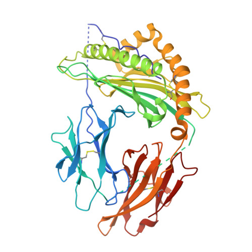
    Type 1 diabetes (T1D) is an autoimmune disease involving T cell-mediated destruction of the insulin-producing beta cells in the pancreatic islets of Langerhans. CD8 + T cells, responding to beta cell peptides presented by class I major histocompatibility complex (MHC) molecules, are important effectors leading to beta cell elimination. Human leukocyte antigen (HLA) B*39:06, B*39:01, and B*38:01 are closely related class I MHC allotypes that nonetheless show differential association with T1D. HLA-B*39:06 is the most predisposing of all HLA class I molecules and is associated with early age at disease onset. B*39:01 is also associated with susceptibility to T1D, but to a lesser extent, though differing from B*39:06 by only two amino acids. HLA-B*38:01, in contrast, is associated with protection from the disease. Upon identifying a peptide that binds to both HLA-B*39:06 and B*39:01, we determined the respective X-ray structures of the two allotypes presenting this peptide to 1.7 Å resolution. The peptide residues available for T cell receptor contact and those serving as anchors were identified. Analysis of the F pocket of HLA-B*39:06 and B*39:01 provided an explanation for the distinct peptide C-terminus preferences of the two allotypes. Structure-based modeling of the protective HLA-B*38:01 suggested a potential reason for its peptide preferences and its reduced propensity to present 8-mer peptides compared to B*39:06. Notably, the three allotypes showed differential binding to peptides derived from beta cell autoantigens. Taken together, our findings should facilitate identification of disease-relevant candidate T cell epitopes and structure-guided therapeutics to interfere with peptide binding.


  • Organizational Affiliation

    Department of Microbiology and Immunology, Albert Einstein College of Medicine, Bronx, NY.


Macromolecules
Find similar proteins by:  (by identity cutoff)  |  3D Structure
Entity ID: 1
MoleculeChains Sequence LengthOrganismDetailsImage
NACHT, LRR and PYD domains-containing protein 2,Beta-2-microglobulin,MHC class I antigen418Homo sapiensMutation(s): 1 
Gene Names: NLRP2NALP2NBS1PAN1PYPAF2B2MCDABP0092HDCMA22PHLA-B
UniProt & NIH Common Fund Data Resources
Find proteins for Q9NX02 (Homo sapiens)
Explore Q9NX02 
Go to UniProtKB:  Q9NX02
PHAROS:  Q9NX02
GTEx:  ENSG00000022556 
Find proteins for R5AK10 (Homo sapiens)
Explore R5AK10 
Go to UniProtKB:  R5AK10
Find proteins for P61769 (Homo sapiens)
Explore P61769 
Go to UniProtKB:  P61769
PHAROS:  P61769
GTEx:  ENSG00000166710 
Entity Groups  
Sequence Clusters30% Identity50% Identity70% Identity90% Identity95% Identity100% Identity
UniProt GroupsQ9NX02R5AK10P61769
Sequence Annotations
Expand
  • Reference Sequence
Small Molecules
Ligands 2 Unique
IDChains Name / Formula / InChI Key2D Diagram3D Interactions
P4G
Query on P4G

Download Ideal Coordinates CCD File 
B [auth A]1-ETHOXY-2-(2-ETHOXYETHOXY)ETHANE
C8 H18 O3
RRQYJINTUHWNHW-UHFFFAOYSA-N
EDO
Query on EDO

Download Ideal Coordinates CCD File 
C [auth A]1,2-ETHANEDIOL
C2 H6 O2
LYCAIKOWRPUZTN-UHFFFAOYSA-N
Experimental Data & Validation

Experimental Data

  • Method: X-RAY DIFFRACTION
  • Resolution: 1.70 Å
  • R-Value Free: 0.232 
  • R-Value Work: 0.202 
  • R-Value Observed: 0.204 
  • Space Group: C 2 2 21
Unit Cell:
Length ( Å )Angle ( ˚ )
a = 75.939α = 90
b = 84.123β = 90
c = 150.421γ = 90
Software Package:
Software NamePurpose
PHENIXrefinement
Aimlessdata scaling
autoPROCdata reduction
PHASERphasing

Structure Validation

View Full Validation Report


Ligand Structure Quality Assessment 


Entry History & Funding Information

Deposition Data


Funding OrganizationLocationGrant Number
National Institutes of Health/National Institute Of Allergy and Infectious Diseases (NIH/NIAID)United StatesR01 AI123730
National Institutes of Health/National Cancer Institute (NIH/NCI)United StatesP30 CA013330

Revision History  (Full details and data files)

  • Version 1.0: 2024-09-04
    Type: Initial release
  • Version 1.1: 2024-10-02
    Changes: Database references