Biochemical and structural insights into a 5' to 3' RNA ligase reveal a potential role in tRNA ligation.
Hu, Y., Lopez, V.A., Xu, H., Pfister, J.P., Song, B., Servage, K.A., Sakurai, M., Jones, B.T., Mendell, J.T., Wang, T., Wu, J., Lambowitz, A.M., Tomchick, D.R., Pawlowski, K., Tagliabracci, V.S.(2024) Proc Natl Acad Sci U S A 121: e2408249121-e2408249121
- PubMed: 39388274 Search on PubMedSearch on PubMed Central
- DOI: https://doi.org/10.1073/pnas.2408249121
- Primary Citation Related Structures:
9C6L, 9C6M - PubMed Abstract:
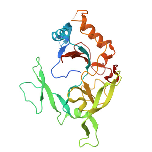
ATP-grasp superfamily enzymes contain a hand-like ATP-binding fold and catalyze a variety of reactions using a similar catalytic mechanism. More than 30 protein families are categorized in this superfamily, and they are involved in a plethora of cellular processes and human diseases. Here, we identify C12orf29 (RLIG1) as an atypical ATP-grasp enzyme that ligates RNA. Human RLIG1 and its homologs autoadenylate on an active site Lys residue as part of a reaction intermediate that specifically ligates RNA halves containing a 5'-phosphate and a 3'-hydroxyl. RLIG1 binds tRNA in cells and can ligate tRNA within the anticodon loop in vitro. Transcriptomic analyses of Rlig1 knockout mice revealed significant alterations in global tRNA levels in the brains of female mice, but not in those of male mice. Furthermore, crystal structures of a RLIG1 homolog from Yasminevirus bound to nucleotides revealed a minimal and atypical RNA ligase fold with a conserved active site architecture that participates in catalysis. Collectively, our results identify RLIG1 as an RNA ligase and suggest its involvement in tRNA biology.
- Department of Molecular Biology, University of Texas Southwestern Medical Center, Dallas, TX 75390.
Organizational Affiliation: