9C5D

E. coli peptidyl-prolyl cis-trans isomerase containing (2S,3S)-4-Fluorovaline


Experimental Data Snapshot

  • Method: X-RAY DIFFRACTION
  • Resolution: 1.30 Å
  • R-Value Free: 0.186 
  • R-Value Work: 0.164 
  • R-Value Observed: 0.165 

Starting Model: experimental
View more details

wwPDB Validation   3D Report Full Report


This is version 1.0 of the entry. See complete history


Literature

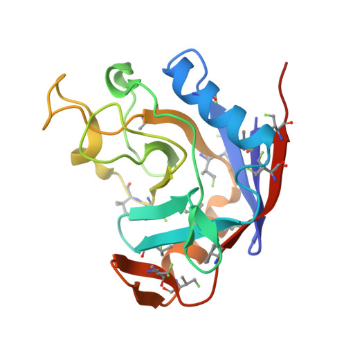
1.3 angstrom Crystal Structure of E. coli Peptidyl-Prolyl Isomerase B with Uniform Substitution of Valine by (2 S ,3 S )-4-Fluorovaline Reveals Structure Conservation and Multiple Staggered Rotamers of CH 2 F Groups.

Frkic, R.L.Tan, Y.J.Maleckis, A.Chilton, N.F.Otting, G.Jackson, C.J.

(2024) Biochemistry 

  • DOI: https://doi.org/10.1021/acs.biochem.4c00345
  • Primary Citation of Related Structures:  
    9C5D

  • PubMed Abstract: 

    (2 S ,3 S )-4-Fluorovaline (FVal) is an analogue of valine, where a single CH 3 group is substituted by a CH 2 F group. In the absence of valine, E. coli valyl-tRNA synthetase uses FVal as a substitute, enabling the production of proteins uniformly labeled with FVal. Here, we describe the production and analysis of E. coli peptidyl-prolyl isomerase B where all 16 valine residues have been replaced by FVal synthesized with a 13 C-labeled CH 2 F group. Although the melting temperature is lower by about 11 °C relative to the wild-type protein, the three-dimensional protein structure is almost completely conserved, as shown by X-ray crystallography. The CH 2 F groups invariably populate staggered rotamers. Most CH 2 F groups populate two different rotamers. The increased space requirement of fluorine versus hydrogen does not prohibit rotamers that position fluorine next to a backbone carbonyl carbon. 19 F NMR spectra show a signal dispersion over 25 ppm. The most high-field shifted 19 F resonances correlate with large 3 J HF coupling constants, confirming the impact of the γ- gauche effect on the signal dispersion. The present work is the second experimental verification of the effect and extends its validity to fluorovaline. The abundance of valine in proteins and structural conservation with FVal renders this valine analogue attractive for probing proteins by 19 F NMR spectroscopy.


  • Organizational Affiliation

    ARC Centre of Excellence for Innovations in Peptide & Protein Science, Research School of Chemistry, Australian National University, Canberra, Australian Capital Territory 2601, Australia.


Macromolecules
Find similar proteins by:  (by identity cutoff)  |  3D Structure
Entity ID: 1
MoleculeChains Sequence LengthOrganismDetailsImage
Peptidyl-prolyl cis-trans isomerase B
A, B, C, D
170Escherichia coliMutation(s): 0 
Gene Names: ppiBb0525JW0514
EC: 5.2.1.8
UniProt
Find proteins for P23869 (Escherichia coli (strain K12))
Explore P23869 
Go to UniProtKB:  P23869
Entity Groups  
Sequence Clusters30% Identity50% Identity70% Identity90% Identity95% Identity100% Identity
UniProt GroupP23869
Sequence Annotations
Expand
  • Reference Sequence
Small Molecules
Ligands 6 Unique
IDChains Name / Formula / InChI Key2D Diagram3D Interactions
XPE
Query on XPE

Download Ideal Coordinates CCD File 
L [auth D]3,6,9,12,15,18,21,24,27-NONAOXANONACOSANE-1,29-DIOL
C20 H42 O11
DTPCFIHYWYONMD-UHFFFAOYSA-N
1PE
Query on 1PE

Download Ideal Coordinates CCD File 
E [auth A]PENTAETHYLENE GLYCOL
C10 H22 O6
JLFNLZLINWHATN-UHFFFAOYSA-N
PGE
Query on PGE

Download Ideal Coordinates CCD File 
G [auth B],
K [auth C]
TRIETHYLENE GLYCOL
C6 H14 O4
ZIBGPFATKBEMQZ-UHFFFAOYSA-N
PEG
Query on PEG

Download Ideal Coordinates CCD File 
I [auth C],
J [auth C]
DI(HYDROXYETHYL)ETHER
C4 H10 O3
MTHSVFCYNBDYFN-UHFFFAOYSA-N
EDO
Query on EDO

Download Ideal Coordinates CCD File 
F [auth A]1,2-ETHANEDIOL
C2 H6 O2
LYCAIKOWRPUZTN-UHFFFAOYSA-N
ACT
Query on ACT

Download Ideal Coordinates CCD File 
H [auth B]ACETATE ION
C2 H3 O2
QTBSBXVTEAMEQO-UHFFFAOYSA-M
Modified Residues  1 Unique
IDChains TypeFormula2D DiagramParent
A1AVU
Query on A1AVU
A, B, C, D
L-PEPTIDE LINKINGC5 H10 F N O2VAL
Experimental Data & Validation

Experimental Data

  • Method: X-RAY DIFFRACTION
  • Resolution: 1.30 Å
  • R-Value Free: 0.186 
  • R-Value Work: 0.164 
  • R-Value Observed: 0.165 
  • Space Group: P 21 21 21
Unit Cell:
Length ( Å )Angle ( ˚ )
a = 79.623α = 90
b = 84.005β = 90
c = 123.432γ = 90
Software Package:
Software NamePurpose
PHENIXrefinement
Aimlessdata scaling
XDSdata reduction
PHASERphasing

Structure Validation

View Full Validation Report


Entry History & Funding Information

Deposition Data


Funding OrganizationLocationGrant Number
Australian Research Council (ARC)AustraliaCE200100012
Australian Research Council (ARC)AustraliaCE200100029

Revision History  (Full details and data files)

  • Version 1.0: 2024-10-09
    Type: Initial release