9C50 | pdb_00009c50

Replacement of a single residue changes the primary specificity of thrombin


Experimental Data Snapshot

  • Method: X-RAY DIFFRACTION
  • Resolution: 2.50 Å
  • R-Value Free: 
    0.245 (Depositor), 0.251 (DCC) 
  • R-Value Work: 
    0.190 (Depositor), 0.196 (DCC) 
  • R-Value Observed: 
    0.193 (Depositor) 

Starting Model: experimental
View more details

wwPDB Validation   3D Report Full Report


This is version 1.1 of the entry. See complete history


Literature

Replacement of a single residue changes the primary specificity of thrombin.

Dei Rossi, A.Deavila, S.Mohammed, B.M.Korolev, S.Di Cera, E.

(2025) J Thromb Haemost 23: 1241-1246

  • DOI: https://doi.org/10.1016/j.jtha.2024.12.024
  • Primary Citation of Related Structures:  
    9C50

  • PubMed Abstract: 

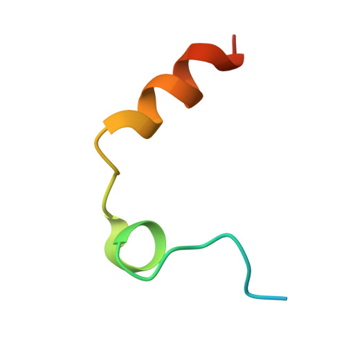
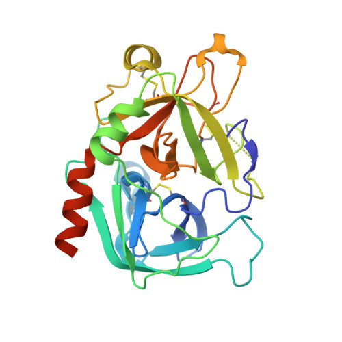
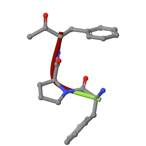
    Thrombin prefers substrates carrying Arg at the site of cleavage (P1) because of the presence of D189 in the primary specificity (S1) pocket but can also cleave substrates carrying Phe at P1. The structural basis of this property is unknown. Solve the X-ray structure of thrombin bound to a ligand carrying Phe at P1 and investigate the effects of replacing D189. X-ray crystallography is used to solve the structure of thrombin bound to the irreversible inhibitor H-D-Phe-Pro-Phe-CH 2 Cl (PPPCK). Residue D189 is mutated to Ala, Lys, Phe and Ser. The X-ray structure of the thrombin-PPPCK complex is solved at 2.5 Å resolution and compared to the structure of thrombin bound to H-D-Phe-Pro-Arg-CH 2 Cl (PPACK). PPPCK binds to thrombin in a conformation similar to that of PPACK, but Phe at P1 makes no contacts with D189. Replacement of D189 with Ala, Lys, Phe or Ser reverses both substrate preference and stability enhancement from Arg to Phe. D189 in the S1 pocket confers thrombin "trypsin-like" specificity for Arg at P1. However, the S1 pocket is wide enough to also enable "chymotrypsin-like" specificity for Phe at P1. Consistent with these structural features, a single amino acid replacement (D189A) switches thrombin specificity from trypsin-like to chymotrypsin-like, converting the substrate preference from H-D-Phe-Pro-Arg-p-nitroanilide to H-D-Phe-Pro-Phe-p-nitroanilide and preferential stability enhancement from PPACK to PPPCK. The observation that thrombin specificity is controlled mainly by a single residue establishes a new paradigm in the field of trypsin-like proteases.


  • Organizational Affiliation
    • Edward A. Doisy Department of Biochemistry and Molecular Biology, Saint Louis University School of Medicine, St. Louis, MO 63104 USA.

Macromolecules
Find similar proteins by:  (by identity cutoff)  |  3D Structure
Entity ID: 1
MoleculeChains Sequence LengthOrganismDetailsImage
Thrombin A-chain
A, C
36Homo sapiensMutation(s): 0 
Gene Names: F2
EC: 3.4.21.5
UniProt & NIH Common Fund Data Resources
Find proteins for P00734 (Homo sapiens)
Explore P00734 
Go to UniProtKB:  P00734
PHAROS:  P00734
GTEx:  ENSG00000180210 
Entity Groups  
Sequence Clusters30% Identity50% Identity70% Identity90% Identity95% Identity100% Identity
UniProt GroupP00734
Sequence Annotations
Expand
  • Reference Sequence
Find similar proteins by:  (by identity cutoff)  |  3D Structure
Entity ID: 2
MoleculeChains Sequence LengthOrganismDetailsImage
Thrombin B-chain
B, D
273Homo sapiensMutation(s): 0 
Gene Names: F2
EC: 3.4.21.5
UniProt & NIH Common Fund Data Resources
Find proteins for P00734 (Homo sapiens)
Explore P00734 
Go to UniProtKB:  P00734
PHAROS:  P00734
GTEx:  ENSG00000180210 
Entity Groups  
Sequence Clusters30% Identity50% Identity70% Identity90% Identity95% Identity100% Identity
UniProt GroupP00734
Sequence Annotations
Expand
  • Reference Sequence

Find similar proteins by:  Sequence   |   3D Structure  

Entity ID: 3
MoleculeChains Sequence LengthOrganismDetailsImage
FPF
E, F
3synthetic constructMutation(s): 0 
Sequence Annotations
Expand
  • Reference Sequence
Small Molecules
Modified Residues  1 Unique
IDChains TypeFormula2D DiagramParent
PCS
Query on PCS
E, F
L-PEPTIDE LINKINGC10 H12 Cl N OPHE
Experimental Data & Validation

Experimental Data

  • Method: X-RAY DIFFRACTION
  • Resolution: 2.50 Å
  • R-Value Free:  0.245 (Depositor), 0.251 (DCC) 
  • R-Value Work:  0.190 (Depositor), 0.196 (DCC) 
  • R-Value Observed: 0.193 (Depositor) 
Space Group: C 1 2 1
Unit Cell:
Length ( Å )Angle ( ˚ )
a = 131.15α = 90
b = 76.732β = 113.77
c = 89.588γ = 90
Software Package:
Software NamePurpose
PHENIXrefinement
HKL-2000data scaling
HKL-2000data reduction
PHASERphasing

Structure Validation

View Full Validation Report


Entry History & Funding Information

Deposition Data


Funding OrganizationLocationGrant Number
National Institutes of Health/National Heart, Lung, and Blood Institute (NIH/NHLBI)United StatesHL049413
National Institutes of Health/National Heart, Lung, and Blood Institute (NIH/NHLBI)United StatesHL139554
National Institutes of Health/National Heart, Lung, and Blood Institute (NIH/NHLBI)United StatesHL147821

Revision History  (Full details and data files)

  • Version 1.0: 2025-01-22
    Type: Initial release
  • Version 1.1: 2025-04-16
    Changes: Database references