9C1U | pdb_00009c1u

Cryo-EM Structure of a Tm1C Fibril


Experimental Data Snapshot

  • Method: ELECTRON MICROSCOPY
  • Resolution: 2.31 Å
  • Aggregation State: HELICAL ARRAY 
  • Reconstruction Method: HELICAL 

wwPDB Validation 3D Report Full Report

Validation slider image for 9C1U

This is version 1.0 of the entry. See complete history

Literature

Cryo-EM and solid state NMR together provide a more comprehensive structural investigation of protein fibrils.

Fonda, B.D.Kato, M.Li, Y.Murray, D.T.

(2024) Protein Sci 33: e5168-e5168

  • DOI: https://doi.org/10.1002/pro.5168
  • Primary Citation Related Structures: 
    9C1U

  • PubMed Abstract: 

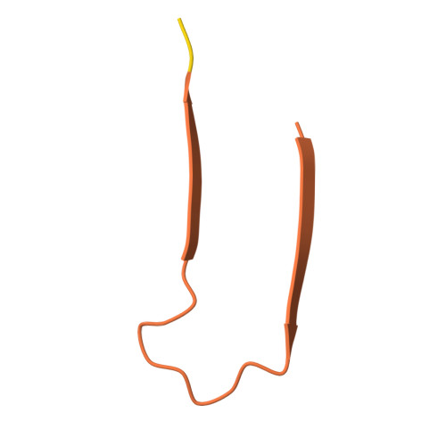
    The tropomyosin 1 isoform I/C C-terminal domain (Tm1-LC) fibril structure is studied jointly with cryogenic electron microscopy (cryo-EM) and solid state nuclear magnetic resonance (NMR). This study demonstrates the complementary nature of these two structural biology techniques. Chemical shift assignments from solid state NMR are used to determine the secondary structure at the level of individual amino acids, which is faithfully seen in cryo-EM reconstructions. Additionally, solid state NMR demonstrates that the region not observed in the reconstructed cryo-EM density is primarily in a highly mobile random coil conformation rather than adopting multiple rigid conformations. Overall, this study illustrates the benefit of investigations combining cryo-EM and solid state NMR to investigate protein fibril structure.


  • Organizational Affiliation
    • Department of Chemistry, University of California, Davis, California, USA.

Macromolecule Content 

  • Total Structure Weight: 89.05 kDa 
  • Atom Count: 2,376 
  • Modeled Residue Count: 312 
  • Deposited Residue Count: 852 
  • Unique protein chains: 1

Macromolecules

Find similar proteins by:|  3D Structure
Entity ID: 1
MoleculeChains  Sequence LengthOrganismDetailsImage
Tropomyosin 1 I/C
A, B, C, D, E
A, B, C, D, E, F, G, H, I, J, K, L
71Drosophila melanogasterMutation(s): 0 
Gene Names: 
UniProt
Find proteins for Q8IG84 (Drosophila melanogaster)
Explore Q8IG84 
Go to UniProtKB:  Q8IG84
Entity Groups
Sequence Clusters30% Identity50% Identity70% Identity90% Identity95% Identity100% Identity
UniProt GroupQ8IG84
Sequence Annotations
Expand
Reference Sequence

Experimental Data & Validation

Experimental Data

  • Method: ELECTRON MICROSCOPY
  • Resolution: 2.31 Å
  • Aggregation State: HELICAL ARRAY 
  • Reconstruction Method: HELICAL 
EM Software:
TaskSoftware PackageVersion
MODEL REFINEMENTPHENIX
RECONSTRUCTIONRELION

Structure Validation

View Full Validation Report


Entry History 

& Funding Information

Deposition Data


Funding OrganizationLocationGrant Number
National Institutes of Health/National Institute of General Medical Sciences (NIH/NIGMS)United StatesR35142892
Cancer Prevention and Research Institute of Texas (CPRIT)United StatesRP220582

Revision History  (Full details and data files)

  • Version 1.0: 2024-10-09
    Type: Initial release