


Domain-swapping promoted by the introduction of a charge in the hydrophobic interior of a protein
Zhang, Y., Schlessman, J.L., Robinson, A.C., Garcia-Moreno E., B., Khangulov, V.S.To be published.
Experimental Data Snapshot
Starting Model: experimental
View more details
Entity ID: 1 | |||||
|---|---|---|---|---|---|
| Molecule | Chains | Sequence Length | Organism | Details | Image |
| Nuclease A | 143 | Staphylococcus aureus | Mutation(s): 4 Gene Names: nuc EC: 3.1.31.1 | 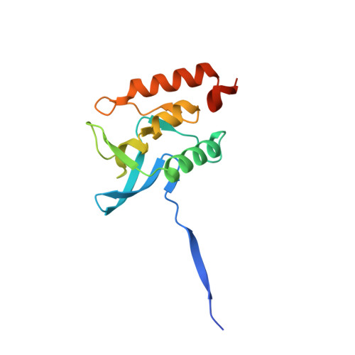 | |
UniProt | |||||
Entity Groups | |||||
| Sequence Clusters | 30% Identity50% Identity70% Identity90% Identity95% Identity100% Identity | ||||
| UniProt Group | P00644 | ||||
Sequence AnnotationsExpand | |||||
Reference Sequence | |||||
| Ligands 2 Unique | |||||
|---|---|---|---|---|---|
| ID | Chains | Name / Formula / InChI Key | 2D Diagram | 3D Interactions | |
| THP Download:Ideal Coordinates CCD File | C [auth A], D [auth B] | THYMIDINE-3',5'-DIPHOSPHATE C10 H16 N2 O11 P2 CSNCBOPUCJOHLS-XLPZGREQSA-N |  | ||
| CA Download:Ideal Coordinates CCD File | E [auth B] | CALCIUM ION Ca BHPQYMZQTOCNFJ-UHFFFAOYSA-N |  | ||
| Length ( Å ) | Angle ( ˚ ) |
|---|---|
| a = 53.183 | α = 90 |
| b = 53.201 | β = 90 |
| c = 107.422 | γ = 90 |
| Software Name | Purpose |
|---|---|
| REFMAC | refinement |
| CrysalisPro | data scaling |
| CrysalisPro | data reduction |
| PHASER | phasing |
| Funding Organization | Location | Grant Number |
|---|---|---|
| National Science Foundation (NSF, United States) | United States | MCB1517378 |
| National Institutes of Health/National Institute of General Medical Sciences (NIH/NIGMS) | United States | GM 061597 |


